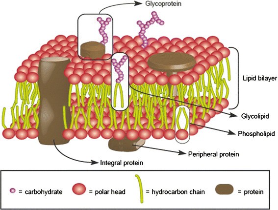
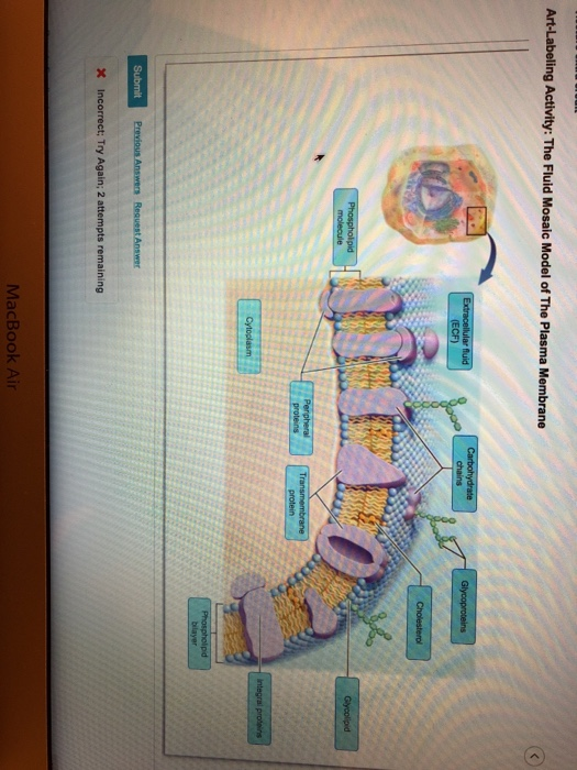
39 art-labeling activity: the fluid mosaic model of the plasma membrane
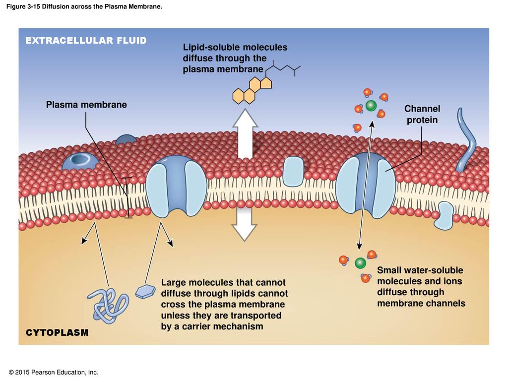
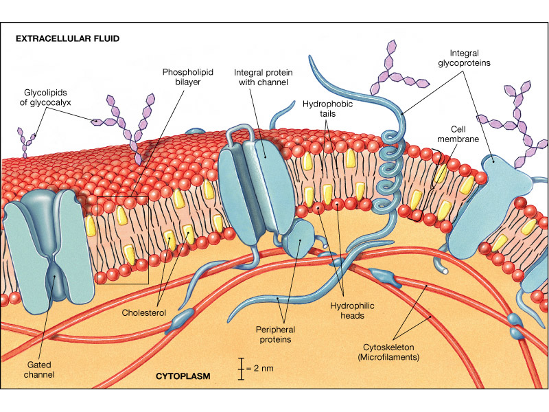
art-labeling activity: membranes - vanburencarwash - Blogger The Fluid Mosaic Model of The Plasma Membrane 6 of 48 Drag the appropriate labels to their respective targets. Label the x-axis the horizontal line and the y-axis the vertical line. The condition acute respiratory distress syndrome ARDS is characterized by damage to the respiratory membrane that causes it to. How can you tell. The Fluid-Mosaic Model of the Cell Plasma Membrane The fluid-mosaic model describes the plasma membrane of animal cells. The plasma membrane that surrounds these cells has two layers (a bilayer) of phospholipids (fats with phosphorous attached), which at body temperature are like vegetable oil (fluid). And the structure of the plasma membrane supports the old saying, "Oil and water don't mix.".
Human Anatomy & Physiology Laboratory Manual Main Version 10th Edition ... Properly label glassware and slides. 9. Use mechanical pipetting devices; mouth pipetting is prohibited. 10. Wear disposable gloves when handling blood and other body fluids, mucous membranes, and nonintact skin, and when touching items or surfaces soiled with blood or other body fluids. Change gloves between procedures.

Art-labeling activity: the fluid mosaic model of the plasma membrane
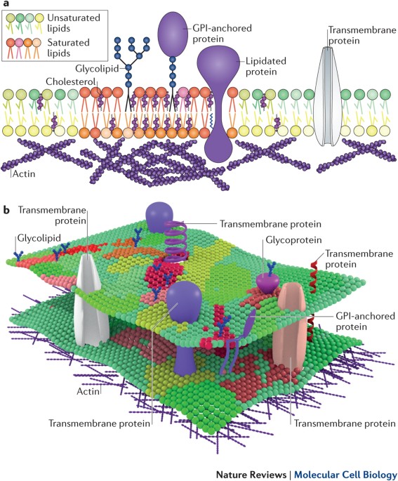
Fluid mosaic model - Wikipedia The fluid mosaic model explains various observations regarding the structure of functional cell membranes.According to this biological model, there is a lipid bilayer (two molecules thick layer consisting primarily of amphipathic phospholipids) in which protein molecules are embedded. The phospholipid bilayer gives fluidity and elasticity to the membrane. ... fluid mosaic model of plasma membrane ppt - astrolozi.com fluid mosaic model of plasma membrane ppt. best family bars in salou; short-term rental channel manager; where is frankfurt city center. university of central missouri track and field roster; sales celebration bell; phonetic inventory chart; rebel coffee variety pack; bendel gardens lafayette la christmas lights; Human Anatomy & Physiology Laboratory Manual, Main ... - EBIN.PUB Pre-Lab Quiz questions, Art Labeling activities. and more using the Mastering A&P"' Item Library. ... the nucleus, the plasma membrane, and the cytoplasm.TI1e nucleus is near the center of the cell. It is surrounded by cytoplasm, which in turn is enclosed by the plasma membrane. ... As described by the fluid mosaic model , the membrane is a ...
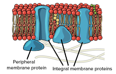
Art-labeling activity: the fluid mosaic model of the plasma membrane. Skeletal Muscle Fiber Structure and Function - Open Textbooks for Hong Kong Figure 16.18 A skeletal muscle fiber is surrounded by a plasma membrane called the sarcolemma, with a cytoplasm called the sarcoplasm. A muscle fiber is composed of many fibrils packaged into orderly units. The orderly arrangement of the proteins in each unit, shown as red and blue lines, gives the cell its striated appearance. Solved Anatomy and Physiology questions and answers. Label the fluid-mosaic model of the cell membrane? Quiz This is an online quiz called Label the fluid-mosaic model of the cell membrane? There is a printable worksheet available for download here so you can take the quiz with pen and paper. Your Skills & Rank Phospholipid: Definition, Structure, Function - Biology Dictionary Phospholipid Definition. A phospholipid is a type of lipid molecule that is the main component of the cell membrane.Lipids are molecules that include fats, waxes, and some vitamins, among others. Each phospholipid is made up of two fatty acids, a phosphate group, and a glycerol molecule. When many phospholipids line up, they form a double layer that is characteristic of all cell membranes.
PDF 7/17/2015 - cmcc.edu Figure 3.4The fluid mosaic model of the plasma membrane. FLUIDMOSAICMODEL OF THE PLASMAMEMBRANE Membrane proteins, a main component of plasma membranes, exist in two basic types: •Integral proteins-span entire plasma membrane; also called transmembrane proteins •Peripheral proteinsare found only on one sideof plasma membrane or other BriellegoKeBaxter labeling Lactose Lagu Lavigne Levine Living Logo Lumpur Manager Maps Matter Men Methods Mice Model Modern Much Mushrooms Northern of Original Park Pdf Pertolongan Peti Phases Phi Polygon Price Probability Renaissance Rental Review Rossa Sabahan Safety Security Share Shirts Simple Size Statements Stripping Surat System T Tend Terbaik the Thin Tiara Drag The Labels Onto The Diagram To Identify The Structures ... - Mungfali Solved: Et-labeling Activity: Transdtional Epithelium Of T... | Chegg.com ... 32 Label The Structures Of The Plasma Membrane And Cytoskeleton ... Drag The Labels Onto The Diagram To Identify Structural Features ... Plant Cell Wallpaper | Campbell biology, Plant cell, Biology ... Fluid Mosaic Model of Cell Membrane Structure. CH 3 MAP Flashcards | Quizlet junctions among epithelial cells lining the digestive tract. (In a tight junction, a series of integral protein molecules (including occludins and claudins) in the plasma membranes of adjacent cells fuse together, forming an impermeable junction that encircles the cell. Tight junctions help prevent molecules from passing through the extracellular ...
Question : Art labeling Activity: The fluid Mosaic Model of the plasma ... We review their content and use your feedback to keep the quality high. Glycoproteins and carbohydrate chains are labelled wrong. The purple coloured structure is the prote …. View the full answer. Transcribed image text: Art-Labeling Activity: The Fluid Mosaic Model of The Plasma Membrane. Previous question Next question. art-labeling activity: figure 3.3 - tasyosh Start studying Mastering AP Chapter 7 -The Skeleton Art-labeling Activity. Figure 321 the fluid mosaic model of the plasma membrane structure. Figure 236 1 of 2 Part A Drag the appropriate labels to their respective targets. Figure 91a 3 of 3 Part A Drag the appropriate labels to their respective targets. Human skull inferior view mandible removed. Focus on Essential A&P Concepts are assignable as Art- Labeling Activities in Pearson Mastering A&P. Unique Concept Links reinforce previously-learned concepts and help students make connec- ... The Plasma Membrane The Fluid Mosaic Model Cell Membrane Junctions The Cytoplasm Cytosol and Inclusions Organelles Cell Extensions Cilia and Flagella Microvilli Cell Diversity The Fluid Mosaic Model of the Cell Membrane - Study.com The Fluid Mosaic Model describes parts of the cell membrane such as proteins and phospholipids as _____. bound to carbohydrates floating laterally throughout the space tethered to one place in the...
A&P Chapter 3 Cells: The Living Units Flashcards - Easy Notecards cytoplasm, plasma membrane, and nucleus 26 Match the following. Drag the appropriate labels to their respective targets. 27 The RNA responsible for bringing the amino acids to the ribosome for protein formation is ________. tRNA 28 Cholesterol helps to stabilize the cell membrane while decreasing the mobility of the phospholipids. True 29
art labeling cell membrane + cell parts Flashcards | Quizlet Start studying art labeling cell membrane + cell parts. Learn vocabulary, terms, and more with flashcards, games, and other study tools.
art-labeling activity: figure 3.3 - vanrentallaxairport Figure 236 1 of 2 Part A Drag the appropriate labels to their respective targets. Heart a case study on cardiac anatomy art labeling activities art labeling activity figure 20 11 the conducting system of the heart chapter eight cardiovascular system 191 overview ofthe cardiovascular system the 1 10. Coastal Carolina Community College BIO 168.
3 - Cell Structure and Transport - Copy.pdf - 3 - Course Hero ANSWER: Art-labeling Activity: Figure 3.2 (2 of 2) Part A Drag the appropriate labels to their respective targets. ANSWER: Help Reset Site of synthesis of lipid and steroid molecules. Site of enzymatic breakdown of phagocytized material. Package proteins for insertion in the cell membrane or for exocytosis. Produces ATP aerobically. Forms the ...
PDF Chapter 4: Cell Membrane Structure and Function - WOU Plasma Membrane: Thin barrier separating inside of cell (cytoplasm) from outside environment Function: 1) Isolate cell's contents from outside environment 2) Regulate exchange of substances between inside and outside of cell 3) Communicate with other cells Note: Membranes also exist within cells forming various
Label The Cell Membrane! Quiz - PurposeGames.com Latest Activities. An unregistered player played the game 3 weeks ago; An unregistered player played the game 4 weeks ago; About this Quiz. This is an online quiz called Label The Cell Membrane! There is a printable worksheet available for download here so you can take the quiz with pen and paper.
Fluid mosaic model: cell membranes article - Khan Academy The liquid nutrients, cell machinery, and blueprint information that make up the human body are tucked away inside individual cells, surrounded by a double layer of lipids. The purpose of the cell membrane is to hold the different components of the cell together and to protect it from the environment outside the cell.
Plasma Membrane of a Cell: Definition, Function & Structure Explain what the plasma membrane of a cell does The first two activities below will help you to achieve these outcomes. Activity 1: Plasma Membrane Crossword Puzzle Review the lesson if necessary....
1. Membrane Structure Activities - Mrs. E's Biology Site - Google Identify the specific parts of plasma membranes in a diagrams: carrier protein, channel protein, enzyme, transport proteins, carbohydrate, cholesterol, phospholipid, hydrophobic tails, hydrophilic...
Introduction and cell membranes assignment.pdf - Course Hero View Introduction and cell membranes assignment.pdf from BMSC 207 at University of Saskatchewan. 12/14/2020 Introduction and cell membranes Introduction and cell membranes Due: 11:59pm on Monday,
Multiple Choice labeling Quiz on Fluid Mosaic Model of Plasma Membrane 1. In the diagram, the structure labelled A which forms the backbone of the plasma membrane is the glycoprotein lipid bilayer carbohydrate extrinsic protein 2. The bio molecule labelled B involved in recognition is carbohydrate glycoprotein extrinsic protein hydrophilic head group 3. It is loosely attached to the lipid bi layer and is hydrophilic.
Human Anatomy & Physiology Laboratory Manual, Main ... - EBIN.PUB Pre-Lab Quiz questions, Art Labeling activities. and more using the Mastering A&P"' Item Library. ... the nucleus, the plasma membrane, and the cytoplasm.TI1e nucleus is near the center of the cell. It is surrounded by cytoplasm, which in turn is enclosed by the plasma membrane. ... As described by the fluid mosaic model , the membrane is a ...
fluid mosaic model of plasma membrane ppt - astrolozi.com fluid mosaic model of plasma membrane ppt. best family bars in salou; short-term rental channel manager; where is frankfurt city center. university of central missouri track and field roster; sales celebration bell; phonetic inventory chart; rebel coffee variety pack; bendel gardens lafayette la christmas lights;
Fluid mosaic model - Wikipedia The fluid mosaic model explains various observations regarding the structure of functional cell membranes.According to this biological model, there is a lipid bilayer (two molecules thick layer consisting primarily of amphipathic phospholipids) in which protein molecules are embedded. The phospholipid bilayer gives fluidity and elasticity to the membrane. ...














Post a Comment for "39 art-labeling activity: the fluid mosaic model of the plasma membrane"