43 transmission electron micrograph labeled
Electron microscope - Wikipedia An electron microscope is a microscope that uses a beam of accelerated electrons as a source of illumination. As the wavelength of an electron can be up to 100,000 times shorter than that of visible light photons, electron microscopes have a higher resolving power than light microscopes and can reveal the structure of smaller objects. A scanning transmission electron … Engineering the Interfacial Microenvironment via Surface … Jul 11, 2022 · The adsorption and activation of CO2 on the electrode interface is a prerequisite and key step for electrocatalytic CO2 reduction reaction (eCO2 RR). Regulating the interfacial microenvironment to promote the adsorption and activation of CO2 is thus of great significance to optimize overall conversion efficiency. Herein, a CO2-philic hydroxyl coordinated ZnO …
Fluorescence microscope - Wikipedia A fluorescence microscope is an optical microscope that uses fluorescence instead of, or in addition to, scattering, reflection, and attenuation or absorption, to study the properties of organic or inorganic substances. "Fluorescence microscope" refers to any microscope that uses fluorescence to generate an image, whether it is a simple set up like an epifluorescence …

Transmission electron micrograph labeled
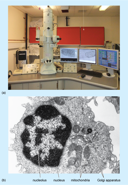
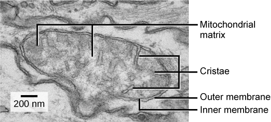
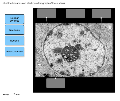
Labeling the Cell Flashcards | Quizlet Label the transmission electron micrograph of the mitochondrion. Label the transmission electron micrograph of the nucleus. membrane bound organelles. golgi apparatus, mitochondrion, lysosome, peroxisome, rough endoplasmic reticulum. nonmembrane bound organelles. ribosomes, centrosome, proteasomes. Transmission electron microscopy - Wikipedia Transmission electron microscopy (TEM) is a microscopy technique in which a beam of electrons is transmitted through a specimen to form an image. The specimen is most often an ultrathin section less than 100 nm thick or a suspension on a grid. An image is formed from the interaction of the electrons with the sample as the beam is transmitted through the specimen. Solved Label the transmission electron micrograph of the - Chegg Expert Answer 100% (4 ratings) Explanation - Mitochondrion is filamentous or globular in shape, occur in variable numbers from a few hundred to few thousands in different cells. It … View the full answer Transcribed image text: Label the transmission electron micrograph of the mitochondrion.
Transmission electron micrograph labeled. Transmission Electron Microscopy - an overview | ScienceDirect … Miroslaw Jonasz, Georges R. Fournier, in Light Scattering by Particles in Water, 2007. 5.7.8 Transmission electron microscopy (TEM). TEM offers a significantly enhanced resolution (0.0001 μm), about one to two orders of magnitude higher than that of the SEM. However, due to the complex process of sample preparation and time-consuming analysis, this technique has … Electron Microscope-Definition, Principle, Types, Uses, Labeled Diagram The electron microscope is placed vertically and has the shape of a tall vacuum column. It consists of the following elements: 1. Electron gun. A heated tungsten filament that produces electrons makes up the electron cannon. 2. Electromagnetic lenses. The condenser lens directs the electron beam to the specimen. Solved Label this transmission electron micrograph of - Chegg Anatomy and Physiology questions and answers Label this transmission electron micrograph of relaxed sarcomeres by clicking and dragging the labels to the correct location Sarcamere 1 band (light) Z disc Mline Aband (dark) H zone microbenotes.com › scanning-electron-microscope-semScanning Electron Microscope (SEM)- Definition, Principle ... Mar 11, 2022 · The first Scanning Electron Microscope was initially made by Mafred von Ardenne in 1937 with an aim to surpass the transmission electron Microscope. He used high-resolution power to scan a small raster using a beam of electrons that were focused on the raster.
Injectable hydrogel microspheres with self-renewable ... - Science Feb 02, 2022 · The transmission electron microscopy (TEM) micrograph of liposomes prepared using a thin-film hydration method revealed a spherical multilamellar vesicle morphology . As shown in Fig. 2B , the liposomes had a positive zeta potential of 45.4 ± 5.6 mV, which would facilitate liposome targeting of negatively charged glycosaminoglycan chains ... Solved Label the transmission electron micrograph of the - Chegg Transcribed image text: Label the transmission electron micrograph of the cell. 0 Nucleus rences Mitochondrion Heterochromatin Peroxisome Vesicle ULAR bumit Click and drag each label into the correct category to indicate whether it pertains to the cytoplasm or the plasma membrane. › articles › s41563/022/01260-ySuperior radiation tolerance via reversible disordering ... May 30, 2022 · A Gatan UltraScan model 994 charge-coupled device camera with an image size of 2,048 × 2,048 pixels in an F30 transmission electron microscope and a Gatan OneView camera (resolution, 4k × 4k) in ... History of HIV/AIDS - Wikipedia In May 1969 16-year-old African-American Robert Rayford died at the St. Louis City Hospital from Kaposi's sarcoma.In 1987 researchers at Tulane University School of Medicine detected a virus closely related or identical to HIV-1 in his preserved blood and tissues. The doctors who worked on his case at the time suspected he was a prostitute or the victim of sexual abuse, though the …
Label This Transmission Electron Micrograph Of A Relaxed ... - Blogger Label this transmission electron micrograph of relaxed sarcomeres by clicking and dragging the labels to the correct location . Label the following image using the terms provided. Note how the sarcomeres are extended to only approximately 120 % . IMG_2132 - FIGURES Label this transmission electron from Solved Label the transmission electron micrograph based on - Chegg Expert Answer nucleus is the house of the genetic material which contains all the h … View the full answer Transcribed image text: Label the transmission electron micrograph based on the hints provided Mitochondrion Heterochromatin Plasma cell Nucleus Rough endoplasmic reticulum Nucleolus Previous question Next question Transmission electron micrograph showing immunogold labeled DipA in the ... Download scientific diagram | Transmission electron micrograph showing immunogold labeled DipA in the outer membrane of the B. burgdorferi Ospless mutant B313. Ultrathin cryosections were prepared ... Transmission electron micrograph of cultured mouse mammary cell labeled ... Download scientific diagram | Transmission electron micrograph of cultured mouse mammary cell labeled as shown in Fig. 3; a thick section was used to increase the length of TMV label included in ...
Cell Micrographs | BioNinja
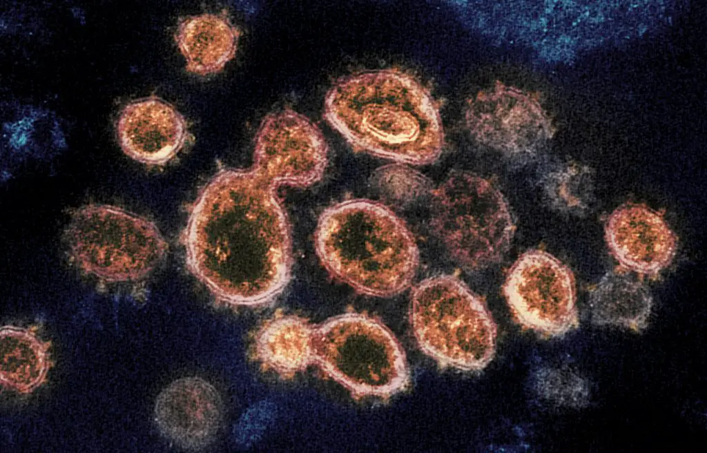
Transmission Electron Microscope (TEM)- Definition, Principle, … May 19, 2022 · Parts of a microscope with functions and labeled diagram; Light Microscope- Definition, Principle, Types, Parts, Labeled Diagram, Magnification ... Transmission electron micrograph of SARS-CoV-2 virus particles, isolated from a patient. Image captured and color-enhanced at the NIAID Integrated Research Facility (IRF) in Fort Detrick, Maryland. ...
Transmission Electron Microscope: Definition, Parts, Working ...
en.wikipedia.org › wiki › Transmission_electronTransmission electron microscopy DNA sequencing - Wikipedia Transmission electron microscopy (TEM) produces high magnification, high resolution images by passing a beam of electrons through a very thin sample. Whereas atomic resolution has been demonstrated with conventional TEM, further improvement in spatial resolution requires correcting the spherical and chromatic aberrations of the microscope lenses.
Transmission Electron Microscope: Definition, Parts, Working ...
Scanning Electron Microscope (SEM)- Definition, Principle, … Mar 11, 2022 · The first Scanning Electron Microscope was initially made by Mafred von Ardenne in 1937 with an aim to surpass the transmission electron Microscope. He used high-resolution power to scan a small raster using a beam of electrons that were focused on the raster. He also aimed at reducing the problems of chromatic aberrations images produced by the …

Transmission Electron Microscope (TEM) - Bioscience Notes
pubs.acs.org › doi › 10Engineering the Interfacial Microenvironment via Surface ... Jul 11, 2022 · The adsorption and activation of CO2 on the electrode interface is a prerequisite and key step for electrocatalytic CO2 reduction reaction (eCO2 RR). Regulating the interfacial microenvironment to promote the adsorption and activation of CO2 is thus of great significance to optimize overall conversion efficiency. Herein, a CO2-philic hydroxyl coordinated ZnO (ZnO–OH) catalyst is fabricated ...
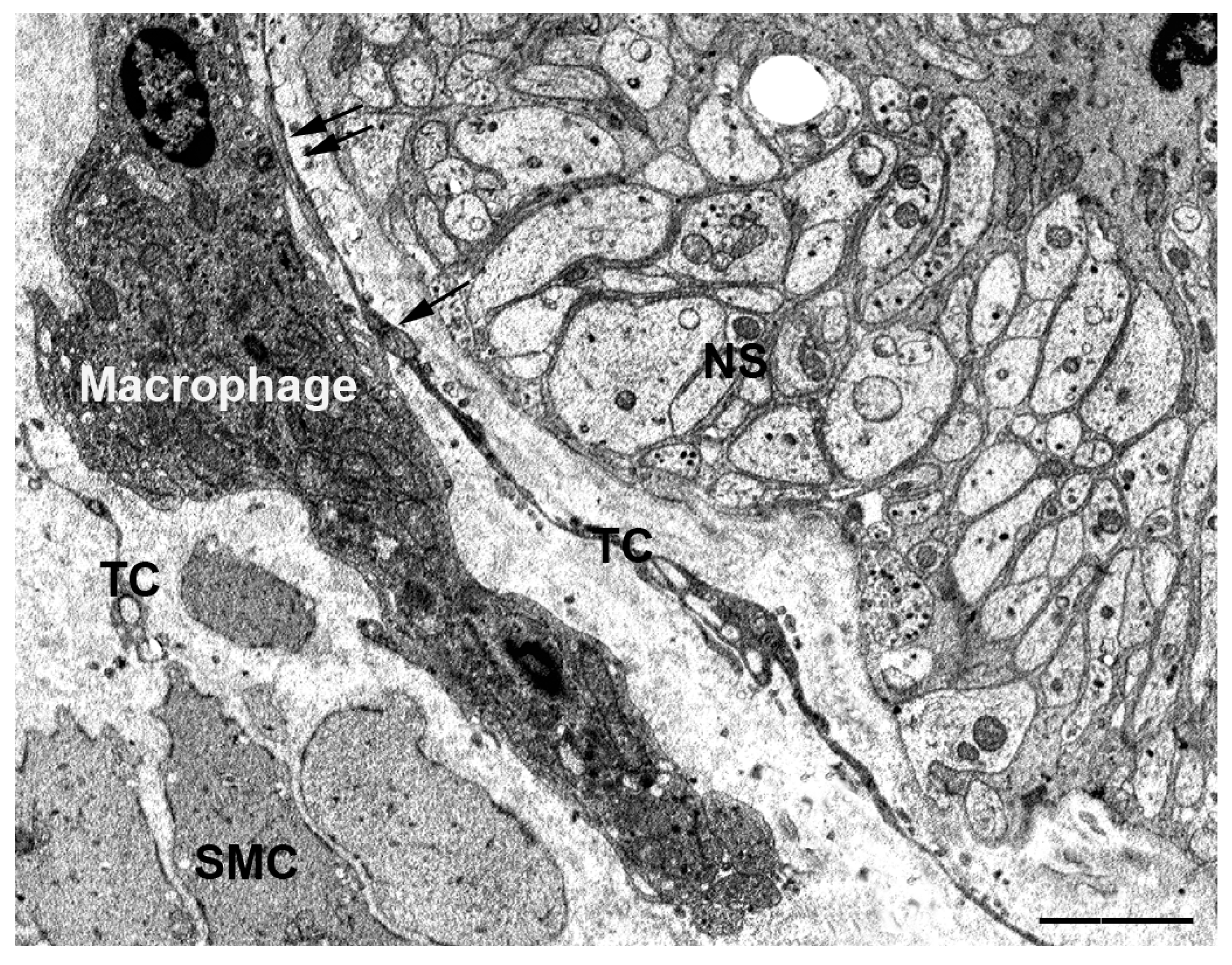
IJMS | Free Full-Text | The Telocytes: Ten Years after Their ...
Transmission Electron Micrograph of transfected HL-1 cells labeled for ... A and B. Single immunogold labeling experiments used 15 nm gold particles to label GFP. A. Immunogold-labeled TMEM43-WT cells. The label (large dot) is associated with the endoplasmic reticulum (arrows), nuclear envelope, and the nucleus (N). B. Immunogold-labeled TMEM43-S358L cells. Immunogold particles (large dot) are confined to bundles of cytoplasmic microfilaments (arrows) and what ...
Electron Microscope Principle, Uses, Types and Images ...
Label This Transmission Electron Micrograph - Kaiden Brown Label this transmission electron micrograph of relaxed sarcomeres by clicking and dragging the labels to the correct location . Transmission electron microscopy (tem) is one of the oldest technologies and still. Molecular labeling for correlative microscopy: Fluorescence microscopy in combination with tem and an ion beam analysis (iba, which ...

Transmission electron microscopy (TEM) of graphene ...
Assignment 6, page 1 Now, it is time to view a chloroplast by transmission electron microscopy. View this transmission electron micrograph of a plant cell, locate a chloroplast and capture the image for labeling.The micrograph is displayed as if using a "virtual electron microscope", so you will need to magnify the image and move to a region that contains the clearest view of chloroplast internal structures.

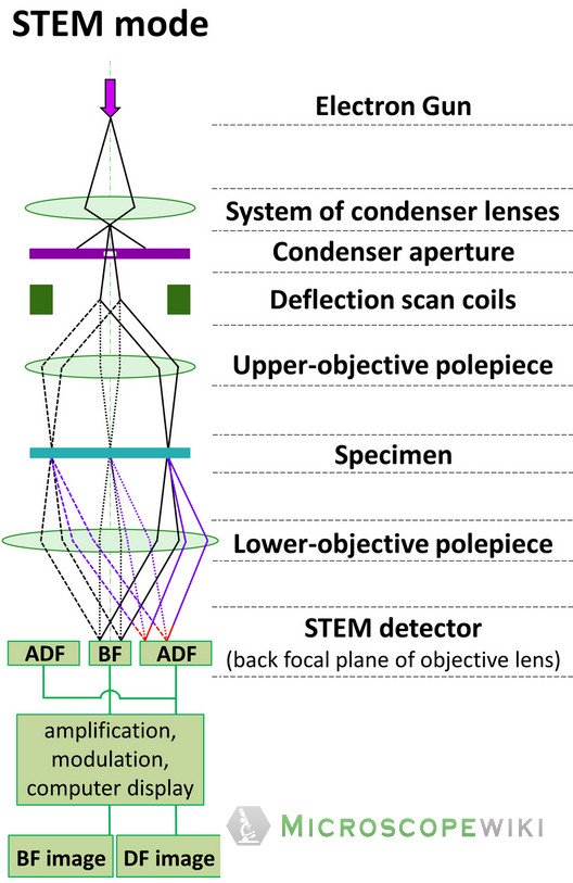
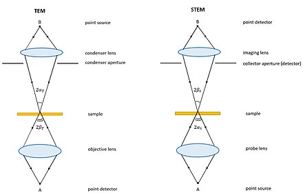
Scanning transmission electron microscopy - Wikipedia
Transmission electron microscopy DNA sequencing - Wikipedia Transmission electron microscopy DNA sequencing is a single-molecule sequencing technology that uses transmission electron microscopy techniques. The method was conceived and developed in the 1960s and 70s, but lost favor when the extent of damage to the sample was recognized. In order for DNA to be clearly visualized under an electron microscope, it must be …

Transmission electron microscope for USPIO-labeled cells ...
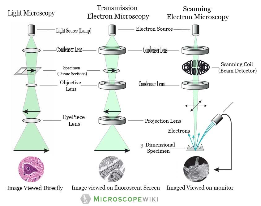
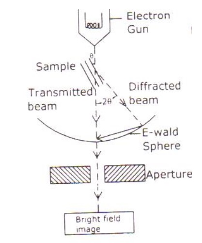
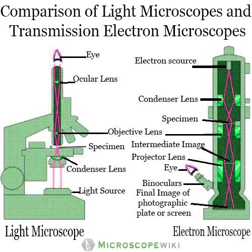
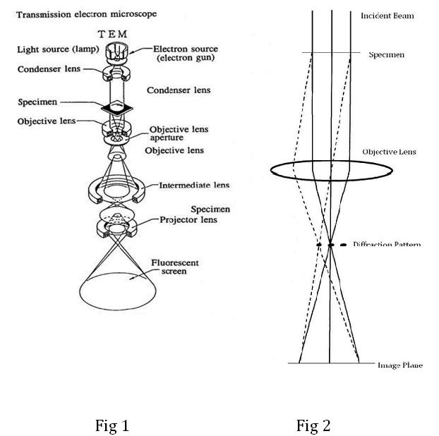
microbenotes.com › transmission-electronTransmission Electron Microscope (TEM)- Definition, Principle ... May 19, 2022 · Principle of Transmission Electron Microscope (TEM) The working principle of the Transmission Electron Microscope (TEM) is similar to the light microscope. The major difference is that light microscopes use light rays to focus and produce an image while the TEM uses a beam of electrons to focus on the specimen, to produce an image.
A tour of the cell: View as single page
› topics › agricultural-andTransmission Electron Microscopy - an overview ... 3.325.5.6 Transmission Electron Microscopy. TEM provides high-resolution imaging and is used for studying small areas or even single mineral platelets selectively. The TEM can be operated at different electron energies (often 100 keV for conventional TEM and 1 MeV for high-resolution imaging). The contrast of the TEM images is dependent on ...
Electron Microscope Principle, Uses, Types and Images ...
en.wikipedia.org › wiki › Electron_microscopeElectron microscope - Wikipedia An electron microscope is a microscope that uses a beam of accelerated electrons as a source of illumination. As the wavelength of an electron can be up to 100,000 times shorter than that of visible light photons, electron microscopes have a higher resolving power than light microscopes and can reveal the structure of smaller objects.

Solved Label the transmission electron micrograph of the ...
Electron Microscope- Definition, Principle, Types, Uses, Labeled Diagram There are two types of electron microscopes, with different operating styles: 1. Transmission Electron Microscope (TEM) The transmission electron microscope is used to view thin specimens through which electrons can pass generating a projection image. The TEM is analogous in many ways to the conventional (compound) light microscope.
Transmission electron microscopy cells hi-res stock ...
Solved Label the transmission electron micrograph of the - Chegg Answer The label is indicated from TOP to BOTTOM Ciliu… View the full answer Transcribed image text : Label the transmission electron micrograph of the cilium.

Scanning Transmission Electron Microscopy - an overview ...
Transmission electron micrograph of a multiple labeled cryosection of ... Download scientific diagram | Transmission electron micrograph of a multiple labeled cryosection of rat skeletal muscle tissue. Section was multiple labeled with anti-alpha-actinin-cAu 6 ...
Transmission Electron microscope - Principle, Construction ...
Superior radiation tolerance via reversible disordering–ordering ... May 30, 2022 · A Gatan UltraScan model 994 charge-coupled device camera with an image size of 2,048 × 2,048 pixels in an F30 transmission electron microscope and a Gatan OneView camera (resolution, 4k × 4k) in ...
Transmission electron microscopy - Wikipedia
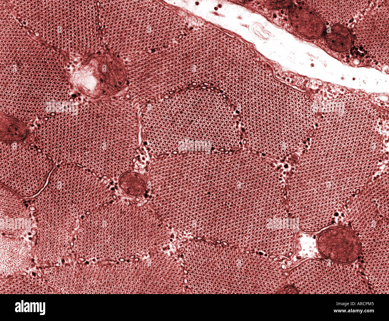
Transmission Electron Microscope (With Diagram) - Biology Discussion The final image in a TEM is known as transmission electron micrograph. The salts of some heavy metals, e.g., lead; osmium, tungsten and uranium are often used for staining. These heavy metal stains are used to increase the contrast between ultra structures and the background.
586 Transmission electron micrograph Images, Stock Photos ...
Microscope Types (with labeled diagrams) and Functions The shorter wavelength of electrons compared to visible light photons helps the observer achieve a very high resolving power compared to normal microscopes thereby aiding observers to see very tiny objects clearly. Electron microscope labeled diagram The different types of electron microscopes are: Transmission Electron Microscope

Introduction to BISC667
Solved Label the transmission electron micrograph of the - Chegg Expert Answer 100% (4 ratings) Explanation - Mitochondrion is filamentous or globular in shape, occur in variable numbers from a few hundred to few thousands in different cells. It … View the full answer Transcribed image text: Label the transmission electron micrograph of the mitochondrion.
FluoroNanogold: Fluorescence and Electron Micrographs of ...
Transmission electron microscopy - Wikipedia Transmission electron microscopy (TEM) is a microscopy technique in which a beam of electrons is transmitted through a specimen to form an image. The specimen is most often an ultrathin section less than 100 nm thick or a suspension on a grid. An image is formed from the interaction of the electrons with the sample as the beam is transmitted through the specimen.

The Transmission Electron Microscope | CCBER
Labeling the Cell Flashcards | Quizlet Label the transmission electron micrograph of the mitochondrion. Label the transmission electron micrograph of the nucleus. membrane bound organelles. golgi apparatus, mitochondrion, lysosome, peroxisome, rough endoplasmic reticulum. nonmembrane bound organelles. ribosomes, centrosome, proteasomes.

Labeling the Cell Flashcards | Quizlet

8.2: Transmission Electron Microscopy - Chemistry LibreTexts

Transmission electron microscopy (TEM). Ten-nanometre gold ...

Transmission Electron Microscope (TEM) - Bioscience Notes
Transmission Electron Microscopy | Central Microscopy ...

Telescope Physics Questions | Questions about Telescope
Electron Microscope Principle, Uses, Types and Images ...

What is Transmission Electron Microscopy?
Transmission electron microscopy cells hi-res stock ...
Preparation of plant cells for transmission electron ...

Transmission Electron Micrograph of transfected HL-1 cells ...
Nuclear envelope. TEM stock image. Image of micrograph ...
Reading: Mitochondria | Biology (Early Release) | | Course Hero
32 Label The Transmission Electron Micrograph Of The Nucleus ...

Transmission electron microscopy DNA sequencing - Wikiwand

Transmission electron micrographs showing phloem cells of ...
What is Transmission Electron Microscopy?
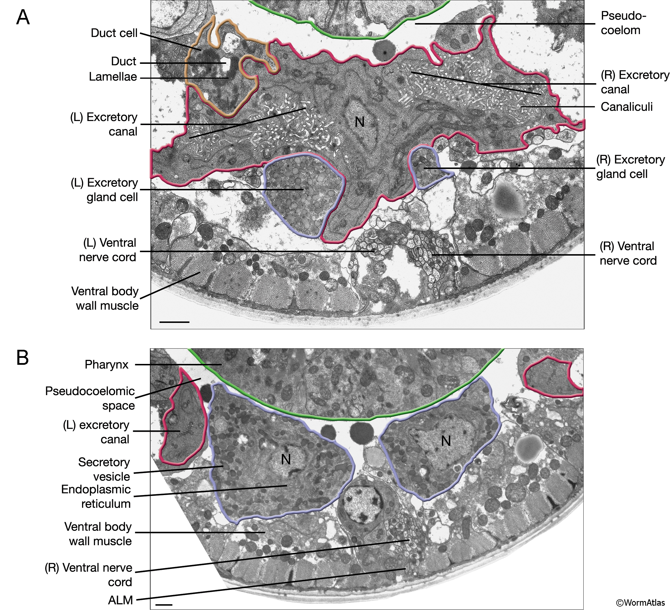
ExcFIG 4 Legend
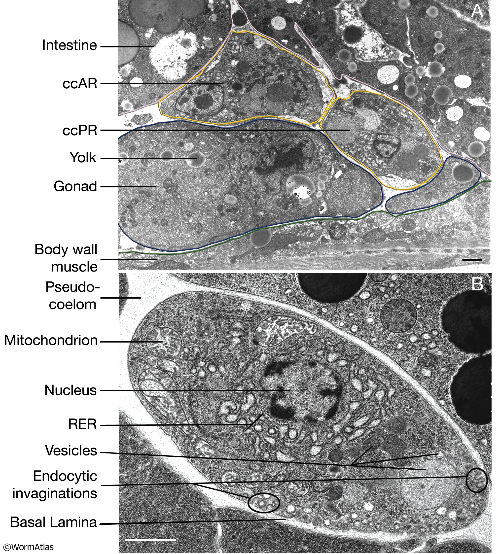
CcFIG 5 Legend

Microscopy

Solved Label the transmission electron micrograph based on ...

A and B) Electron micrograph of a cell labeled for/5-tubulin ...
Transmission Electron Microscopy (TEM)
Post a Comment for "43 transmission electron micrograph labeled"