41 label the parts of the microscope
ECLIPSE Ti2 Series | Inverted Microscopes | Nikon Microscope Products ... A collection of built-in sensors detects and relays status information for a variety of components in the microscope. All of the status information is recorded in the metadata when you acquire images with a computer, so you can easily recall acquisition conditions and/or check for configuration errors. Microscope Labeling Game - PurposeGames.com About this Quiz. This is an online quiz called Microscope Labeling Game. There is a printable worksheet available for download here so you can take the quiz with pen and paper.. This quiz has tags. Click on the tags below to find other quizzes on the same subject.
Optical biosensing through a toy microscope over a surface 'rainbow' chip The spatial location of the dark bar is determined by the incident wavelength, that is, different incident wavelength will result in different dark bars at different positions within a 30 μm × 64...

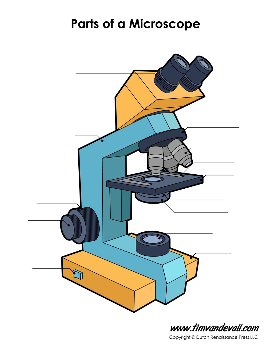
Label the parts of the microscope
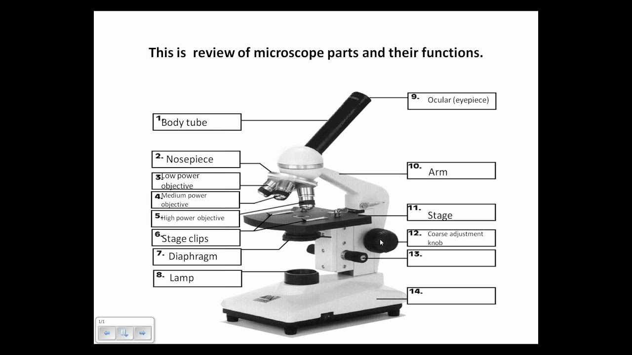
Microscope Activity Worksheet [ZPA1C5] section 4 - microscopes!1 of 5 study the following table, which describes features of the compound light microscope and the function of each light microscope (lm)-employs visible light to detect small objects (uses focused light) lm description and what it is used on-2 lenses *ocular lens and objective lens-fluorescent dyes can be used to … Metaphase - Genome.gov Normally, individual chromosomes are spread out in the cell nucleus. During metaphase, the nucleus dissolves and the cell's chromosomes condense and move together, aligning in the center of the dividing cell. At this stage, the chromosomes are distinguishable when viewed through a microscope. Parts of a Microscope with Their Functions - Microbe Online Parts of Compound Microscope Illuminator (Light Source) Diaphragm (Iris) Condenser Aperture Stage Objective lens Body Tube Ocular Lens (eye-piece) Coarse and Fine Adjustment Knob Arm Base Microscope Worksheet The Light Microscope Light microscopes are used to examine cells at relatively low magnifications.
Label the parts of the microscope. Bruins Facing Tough Roster Decision With Studnicka Ever since he was drafted 53rd overall in the second round of the 2017 Entry Draft, the microscope has been on Studnicka. He has been labeled as the center in waiting for when it becomes reality... Microscope Imaging Software | Products | Leica Microsystems 23/08/2021 · Microscope Imaging Software. Microscope imaging software from Leica Microsystems combines microscope, digital camera and accessories into one fully integrated solution. With an intuitive user interface and straightforward navigation, it guides the user through any workflow, whether fast image acquisition or sophisticated expert analysis. A ... Groundbreaking Method "Starves" Highly-Lethal Cancer Tumors of Energy ... Under the microscope, we found that activated astrocytes surrounded glioblastoma tumors. Based on this observation, we set out to investigate the role of astrocytes in glioblastoma tumor growth." Using an animal model, in which they could eliminate active astrocytes around the tumor, the researchers found that in the presence of astrocytes ... fossil | Definition, Types, Examples, & Facts | Britannica fossil, remnant, impression, or trace of an animal or plant of a past geologic age that has been preserved in Earth's crust. The complex of data recorded in fossils worldwide—known as the fossil record—is the primary source of information about the history of life on Earth. Only a small fraction of ancient organisms are preserved as fossils, and usually only organisms that have a solid ...
› game › microscope-labelingMicroscope Labeling Game - PurposeGames.com This is an online quiz called Microscope Labeling Game. There is a printable worksheet available for download here so you can take the quiz with pen and paper. This quiz has tags. Click on the tags below to find other quizzes on the same subject. Slit Lamp - FPnotebook.com An eye exam using an instrument that combines a low-power microscope with a light source that makes a narrow beam of light. The instrument may be used to examine the retina, optic nerve, and other parts of the eye. Examination of the anterior segment of the eye using a medical instrument called a slit lamp. rsscience.com › stereo-microscopeParts of Stereo Microscope (Dissecting microscope) – labeled ... Labeled part diagram of a stereo microscope Major structural parts of a stereo microscope. There are three major structural parts of a stereo microscope. The viewing Head includes the upper part of the microscope, which houses the most critical optical components, including the eyepiece, objective lens, and light source of the microscope. University of Arizona scientists go to great lengths to study microbes The chance for a closer look at the tiny, invisible creatures that inhabit nearly every square inch of the Earth's surface. Microbes. In two tablespoons of soil, Carini says, there can be as ...
Parts of a Dog - External Body Features of Dogs with Diagram Parts of the dog's head In the head of a dog, you will find the cranium (skull), occiput, stop, nose, mouth, muzzle, lip, flew, and cheek. Again, you might also identify the ear, eye, foreface, jaw point, and teeth from the dog's head area. Now, let's know the details of these features from the head area of a dog. Virtual Labs: Using the Microscope - GameUp - BrainPOP. In this free online science interactive, students learn the procedures for operating a compound optical light microscope as they would use in a science lab. bVX0-zncj9qJ3G1_r18rkIpQL02X-Oi6tWViR4g4-vwDVmU50WZA-4bRZMjM2TXmc88PAkJ1g0jIembnEbM Karyotype - Genome.gov The typical human karyotype contains 22 pairs of autosomal chromosomes and one pair of sex chromosomes. The most common karyotypes for a female contain two X chromosomes and are denoted for the sex XX. Males usually have both an X and a Y chromosome, denoted for the sex XY. Amalia Dutra, Ph.D. Director Cytogenetics and Microscopy Core › game › label-the-model-humanLabel the Model Human Cell Quiz - PurposeGames.com Three Letter Body Parts 8p Type-the-Answer. ... This is an online quiz called Label the Model Human Cell . ... Compound Light Microscope 17p Shape Quiz.
Label the light microscope | Teaching Resources
NFL community rips Dolphins over Tua Tagovailoa injury Part of the league's protocols that would result in a "no-go" for a return to the field include gross motor instability. ... the microscope on the Dolphins and their decision making has only ...
Parts of the Microscope Labeling Activity!
5 White Blood Cells Types and Their Functions - New Health Advisor There are two different kinds of white blood cells and each looks different from one another under the microscope. These include granulocytes and agranulocytes. Granulocytes have visible granules or grains inside the cells that have different cell functions. Types of granulocytes include basophils, neutrophils, and eosinophils.
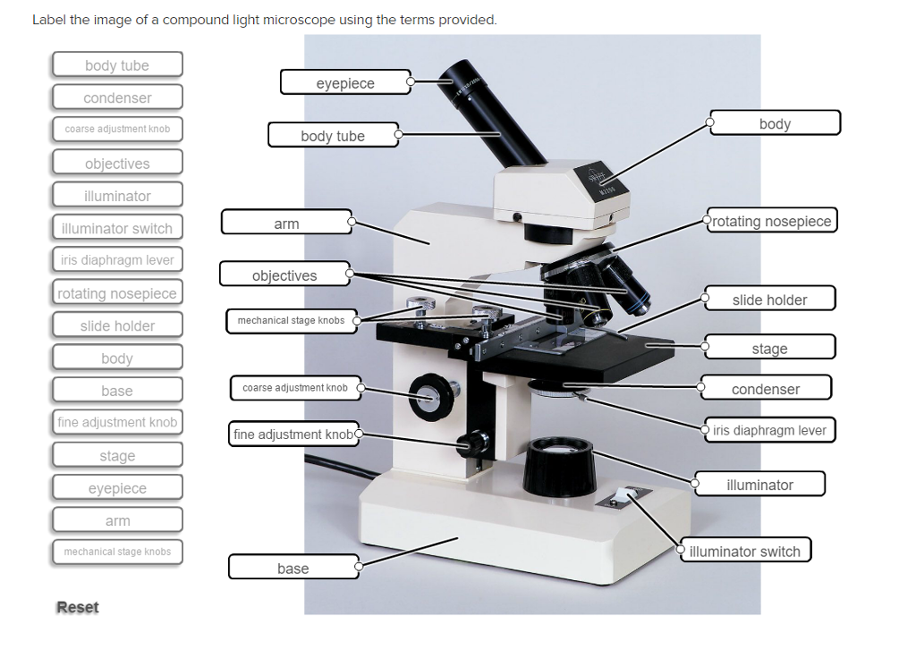
Solved Label the image of a compound light microscope using ...
Science Snapshot: More Fun Than a Barrel of Monkey Cells T his year's 2nd place winner of the Nikon Small World in Motion competition went to Christophe Leterrier of Aix Marseille Université for a time-lapse of cultured monkey cells. Leterrier's confocal microscope magnified the cells 60X, and the researcher distilled 12 hours of footage into a 12-second video that shows how dynamic cells (plasma membrane in orange) and DNA (blue) can be.

Parts of a Compound Microscope and Their Functions
Label the Model Human Cell Quiz - PurposeGames.com Three Letter Body Parts 8p Type-the-Answer. Time Zones of the USA 6p Image Quiz. Who's the oldest? 10p Order Quiz • • • Play Again. Surprise Me! Next Game » For You; Badges (66) Playlists (7) Tournaments (37) AI Stream The more you play, the more accurate suggestions for you. Your Streams. Loading ... Cities by Landmarks 11p Image Quiz. The worlds tallest buildings 9p …

Parts of the Microscope with Labeling (also Free Printouts ...
Microscope Labelled Diagram Gcse Micropedia - Otosection Surface Studio vs iMac - Which Should You Pick? 5 Ways to Connect Wireless Headphones to TV. Design

Compound Microscope: Parts of Compound Microscope
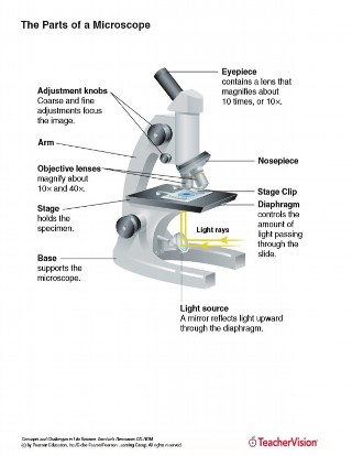
Light Microscope (Theory) - Amrita Vishwa Vidyapeetham The modern compound microscope consists of two lens system, the objective and the ocular or eye piece. The first magnified image obtained with objective lens, is again magnified by the eye piece to give a virtual inverted image. The total magnification the product of the magnifications of two lens systems. Parts of a Microscope
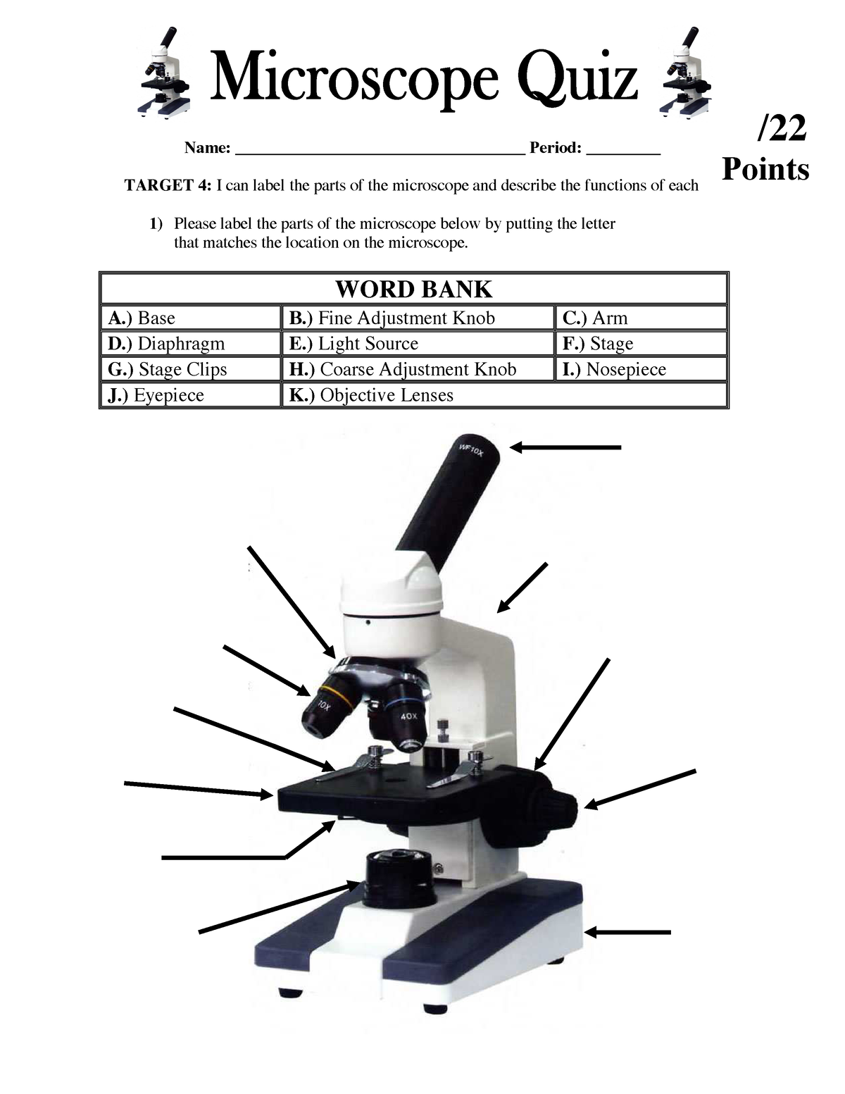
Parts of the Microscope Quiz Lesson 2 - Name: Period: TARGET ...
UD Virtual Compound Microscope - University of Delaware ©University of Delaware. This work is licensed under a Creative Commons Attribution-NonCommercial-NoDerivs 2.5 License.Creative Commons Attribution-NonCommercial-NoDerivs 2.5 License.

Compound Microscope Parts – Labeled Diagram and their ...
Parts of a microscope with functions and labeled diagram 17/09/2022 · Thank you very much it really helped me with my science home work since i in 8th grade and this my home work to draw a microscope label all the parts and the function thank may the holy father of holy spirits bless you and give more wisdom thanks love you all keep up the good work and thank you again bye. Reply

Microscope Label Teaching Resources | Teachers Pay Teachers
Light Microscope (Assignment) - Amrita Vishwa Vidyapeetham The first experiment i.e.; knowing the parts of a microscope is a self explanatory animation. Instructor's can choose this animation in the class before detailing the parts of a microscope. To illustrate the working of a microscope, follow the instructions in the simulator. Students Assignment
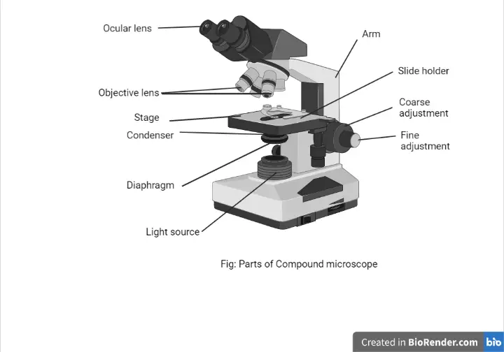
Parts of a Microscope with Their Functions – Microbe Online
Microscope slide - Wikipedia A microscope slide is a thin flat piece of glass, typically 75 by 26 mm (3 by 1 inches) and about 1 mm thick, used to hold objects for examination under a microscope.Typically the object is mounted (secured) on the slide, and then both are inserted together in the microscope for viewing. This arrangement allows several slide-mounted objects to be quickly inserted and …

16 Basic Parts of Microscope, Function, Names & Labeled Diagram
Autoclave: Principle, Procedure, Types, Uses - Microbe Online The usual procedure is to heat at 1.1 kilograms/square centimeter (kg/cm 2) [15 pounds/square inch (lb/in 2 )] steam pressure, which yields a temperature of 121°C. At 121°C, the time of autoclaving to achieve sterilization is generally considered to be 15-20 min, depending on the volume of the load. To make sure, sterilization is successful ...
Microscope parts
NCBioNetwork.org | Life Science Education, Training, and Laboratory ... Microscope Basics eLearning ; 1 of 53 > BioWork The BioWork certificate program will teach you the foundational skills you need to begin a career as a process technician for a biotechnology, pharmaceutical, or chemical manufacturing company. Process technicians are responsible for the production of chemical and pharmaceutical products.

School | Reading worksheets, Microscope parts, Biology lessons
microbenotes.com › parts-of-a-microscopeParts of a microscope with functions and labeled diagram Sep 17, 2022 · Figure: Diagram of parts of a microscope. There are three structural parts of the microscope i.e. head, base, and arm. Head – This is also known as the body. It carries the optical parts in the upper part of the microscope. Base – It acts as microscopes support. It also carries microscopic illuminators.

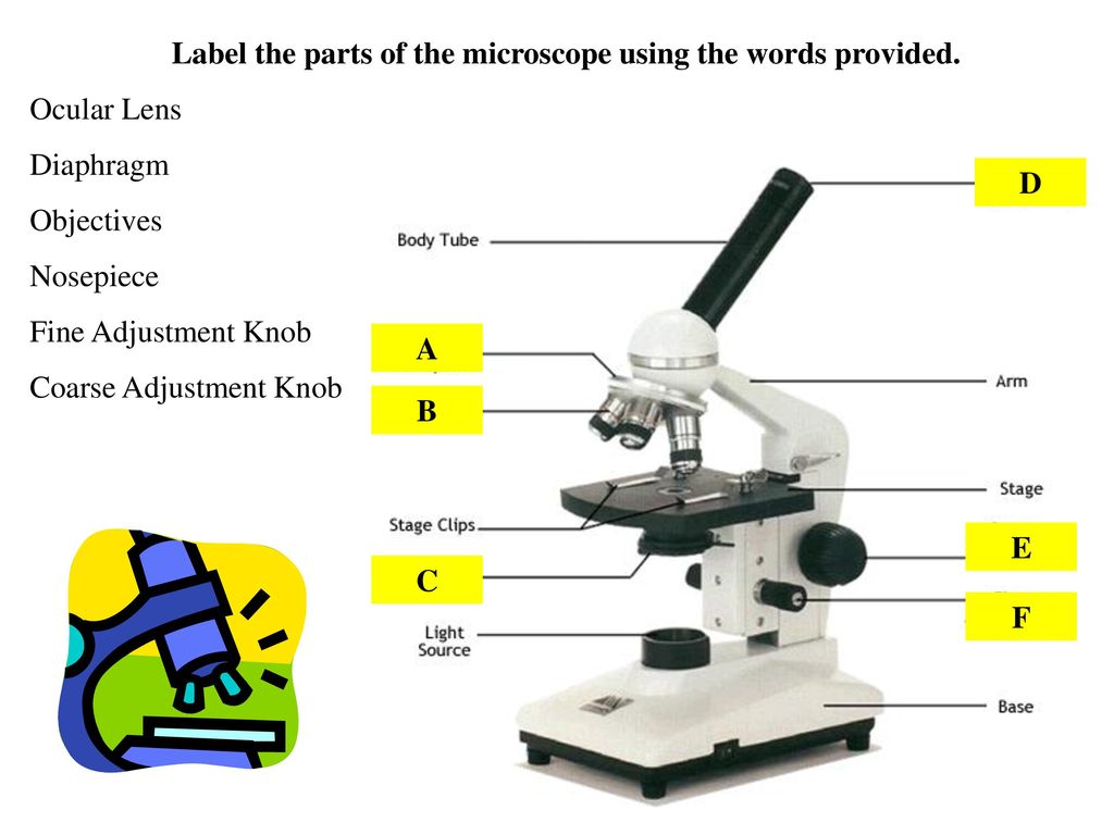
Learning Task 3: Label Me!Label The Parts of the Microscope ...
Smoker's Lung Pictures: Smokers' Lungs vs. Healthy Lungs - MedicineNet Figure 1 is a diagram showing the main parts of the airway and lung. The airway consists of the oral and nasal cavities, which connect to the voice box (larynx), which connects to the windpipe (trachea). Note in the diagram that the windpipe splits into two air passages called bronchi, one going to each lung (right and left main bronchi).

Microscope Components - Science Quiz
Mr. Jones's Science Class Earth, Moon, & Sun System (PPT.) Seasons Interactive (Online Activity) Moon Phases - Introductory Activity. Modeling the Phases of the Moon. Problems in Space (Online Activity) Lunar & Solar Eclipses - Webquest.
Parts of a Light Microscope Activity | Labeling Task
Parts of Stereo Microscope (Dissecting microscope) – labeled … Optical parts of a stereo microscope work together to magnify and produce a 3-D image of the specimens. These parts include: Eyepieces. The eyepiece (or ocular lens) is the lens part at the top of a microscope that the viewer looks through. Typically, standard eyepieces for a dissecting microscope have a magnifying power of 10x. Optional eyepieces of varying powers are …
The Parts of a Microscope (Labeled) Printable Printable (6th ...
Diagram of a Compound Microscope - Biology Discussion ADVERTISEMENTS: In this article we will discuss about:- 1. Essential Parts of Compound Microscope 2. Magnification of the Image of the Object by Compound Microscope 3. Resolution Power 4. Method for Studying Microbes 5. Measurement of the Size of Objects. Essential Parts of Compound Microscope: The essential parts of usually used monocular compound …

Biology label part of microscope
› games › virtuallabsusingthemicroscopeVirtual Labs: Using the Microscope - GameUp - BrainPOP. In this free online science interactive, students learn the procedures for operating a compound optical light microscope as they would use in a science lab. bVX0-zncj9qJ3G1_r18rkIpQL02X-Oi6tWViR4g4-vwDVmU50WZA-4bRZMjM2TXmc88PAkJ1g0jIembnEbM

B. ExercisesExercise 1: REMEMBER ME? THEN WRITE MY NAME ...
11 Different Types of Microscopes (With Pictures) - Optics Mag The 11 Types of Microscopes: 1. Light Microscopes The most common type of microscope you're likely to come across, these microscopes rely on lenses and light to illuminate a specimen for optimal image-gathering. They can be used for viewing living cells, insects, for performing dissections, or for clinical blood and tissue assessment. 2.

Parts of a microscope with functions and labeled diagram
www1.udel.edu › biology › ketchamUD Virtual Compound Microscope - University of Delaware ©University of Delaware. This work is licensed under a Creative Commons Attribution-NonCommercial-NoDerivs 2.5 License.Creative Commons Attribution-NonCommercial-NoDerivs 2

Label the Parts of the Microscope - Brainly.ph
Spinal Cord Cross Section | New Health Advisor The gray matter is the core and ends up to be four projections that are known as horns. At the back are two dorsal horns and away from the back are two ventral horns. The gray matter found here consists of interneurons, neurons, and glial cells, all part of the central nervous system.
Microscope Bundle!! - Parts of a Microscope Unit Activities | TpT
› iet › microscopeVirtual Microscope - NCBioNetwork.org Lesson Description BioNetwork’s Virtual Microscope is the first fully interactive 3D scope - it’s a great practice tool to prepare you for working in a science lab. Explore topics on usage, care, terminology and then interact with a fully functional, virtual microscope.
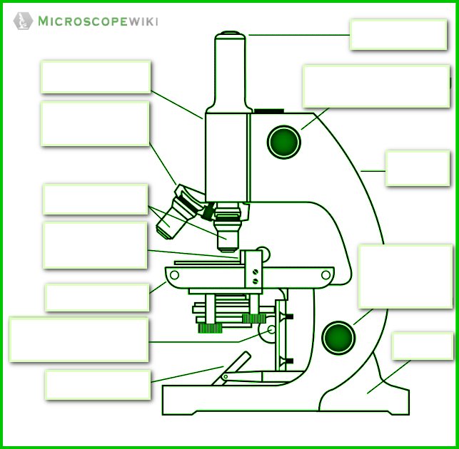
Microscope Diagram Labeled, Unlabeled and Blank | Parts of a ...
The Harms of Cancer Screening They Don't Warn You About Yes, harms. Decades of screening by ever-more perceptive advanced technologies have taught us that not all cancers kill. In fact, many tiny early cancers that screening can now detect never spread ...

Label the diagram of the microscope and explain the role of ...
Accurate and fast identification of minimally prepared bacteria ... The spectral transformer (ST) consists of an optional positional embedding layer, followed by a dropout layer. The next layer is a transformer-encoder block that sequentially contains, layer...

Microscope World Blog: Labeling the Parts of the Microscope
Virtual Microscope - NCBioNetwork.org Lesson Description BioNetwork’s Virtual Microscope is the first fully interactive 3D scope - it’s a great practice tool to prepare you for working in a science lab. Explore topics on usage, care, terminology and then interact with a fully functional, virtual microscope. When you are ready, challenge your knowledge in the testing section to see what you have learned.

Biology label part of microscope
Olympus U-Tru Microscope Side Camera - acmerevival.com Olympus U-Tru Microscope Side Camera. 1 units left at this price. what is this? 16 people are currently viewing this device. $1,421.00. Acme Revival's warranty coverage protects your device in-case of malfunction/defect, or in-case of accidents such as drops, or water damage.

Label-microscope.docx - Label parts of the Microscope ...
Parts of a Microscope with Their Functions - Microbe Online Parts of Compound Microscope Illuminator (Light Source) Diaphragm (Iris) Condenser Aperture Stage Objective lens Body Tube Ocular Lens (eye-piece) Coarse and Fine Adjustment Knob Arm Base Microscope Worksheet The Light Microscope Light microscopes are used to examine cells at relatively low magnifications.
(159).jpg)
Microscope Quiz: How Much You Know About Microscope Parts And ...
Metaphase - Genome.gov Normally, individual chromosomes are spread out in the cell nucleus. During metaphase, the nucleus dissolves and the cell's chromosomes condense and move together, aligning in the center of the dividing cell. At this stage, the chromosomes are distinguishable when viewed through a microscope.

This is a common compound microscope Label its parts class 11 ...
Microscope Activity Worksheet [ZPA1C5] section 4 - microscopes!1 of 5 study the following table, which describes features of the compound light microscope and the function of each light microscope (lm)-employs visible light to detect small objects (uses focused light) lm description and what it is used on-2 lenses *ocular lens and objective lens-fluorescent dyes can be used to …

Microscope Parts worksheet

label microscope diagram | Charts | Microscope, Anatomy bones ...
Name Date Sci STANDARD MICROSCOPE DIAGRAM Label only the ...
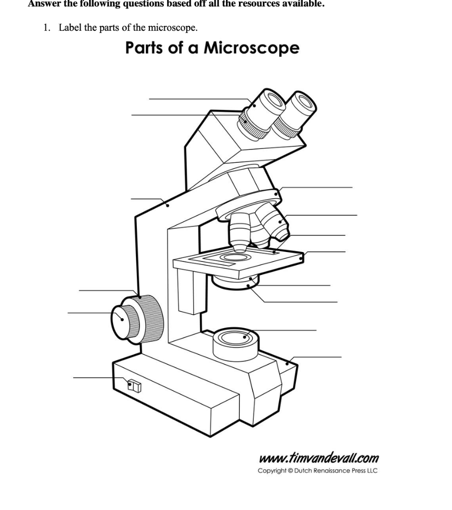
Solved Answer the following questions based off all the ...
Microscope Review.wmv - YouTube
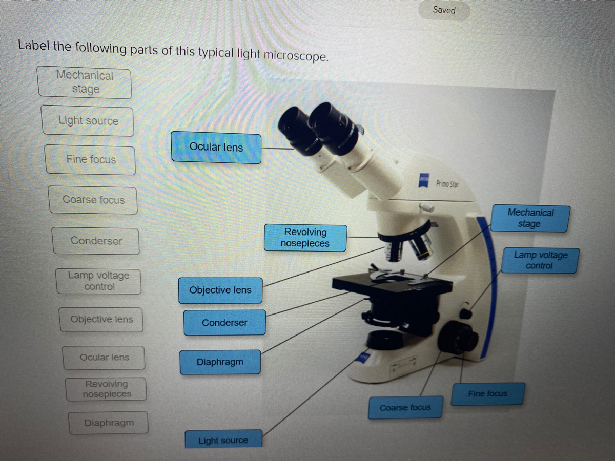
Answered: Saved Label the following parts of this… | bartleby

Compound Microscope Parts – Labeled Diagram and their ...
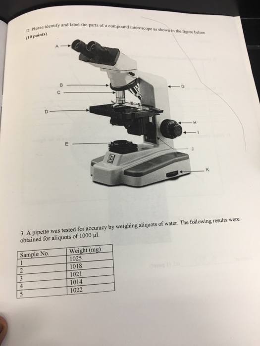
Solved Identify and label the parts of a compound microscope ...
Challenge #1 Parts of the Microscope - ppt download
Simple Microscope - Diagram (Parts labelled), Principle ...

This is a common compound microscope. Label its parts from A ...

Label the parts and functions of the microscope no. 1-16 ...
Post a Comment for "41 label the parts of the microscope"