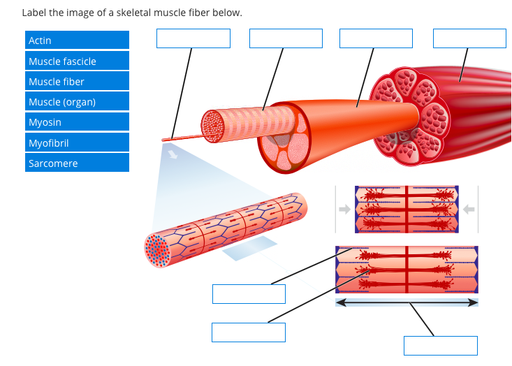
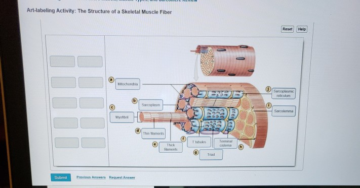
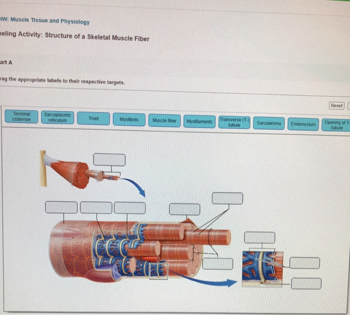
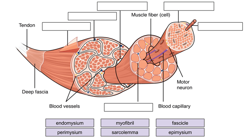
45 label the structures of a skeletal muscle fiber
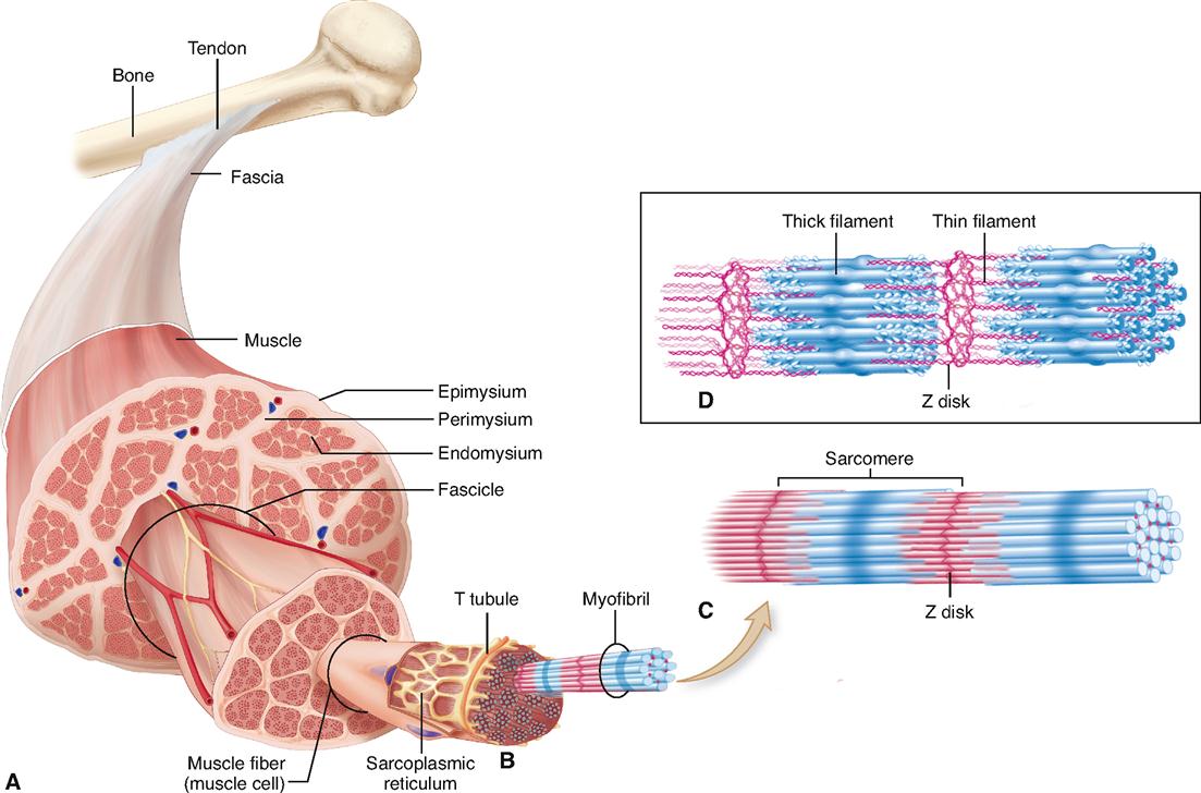
Label structure of skeletal muscle Diagram | Quizlet Label structure of skeletal muscle 4.0 (5 reviews) + − Learn Test Match Created by danielaaaa04 Teacher Terms in this set (8) Term myofibrils Location Term sarcoplasmis reticulum Location Term sarcolemma Location Term epimysium Location Term perimysium Location Term endomysium Location Term fascicle Location Term muscle fiber Location Muscle Fibers: Anatomy, Function, and More - Healthline Skeletal muscle fibers are classified into two types: type 1 and type 2. Type 2 is further broken down into subtypes. Type 1. These fibers utilize oxygen to generate energy for movement. Type...
unlabeled skeletal muscle anatomy Skeletal structure fibers function fascicle quizlet myofibrils endomysium perimysium exercising fascicles contraction fascia myofibril surrounded exercise labeling 1001. Skeletal muscle fiber model quiz. Muscles facial face risorius frontalis many smile skeleton skull used spasms building there wrinkles under eye aerobics build exercises ...
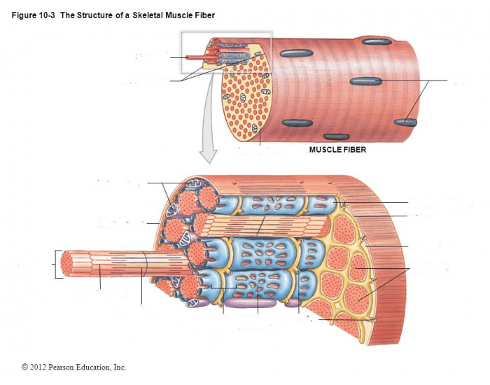
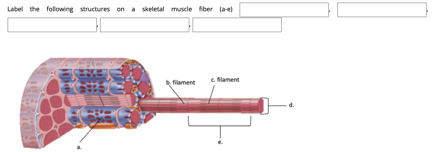
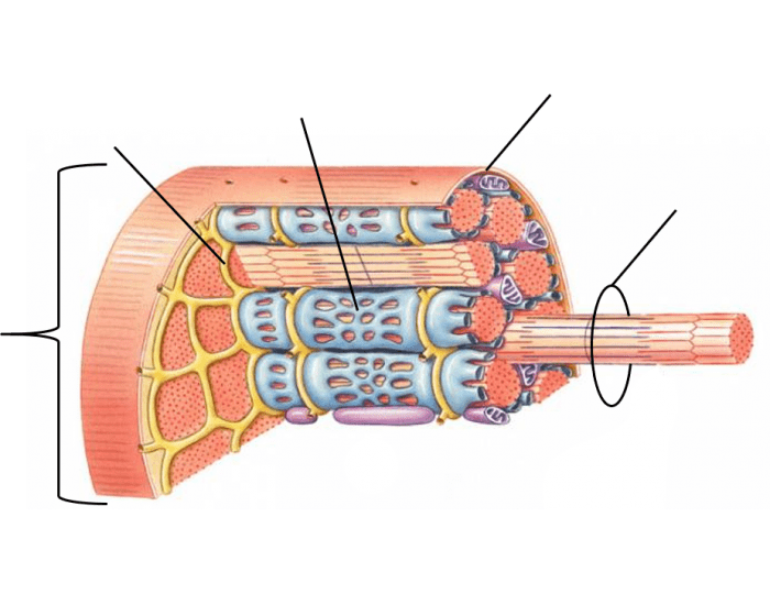
Label the structures of a skeletal muscle fiber
IJMS | Free Full-Text | Comprehensive Analysis of Long Noncoding RNA ... Skeletal muscle is a heterogeneous tissue, composed of different types of muscle fibers, with distinct contractile and metabolic characteristics [].Myosin heavy chain (MyHC) is the major contractile protein of skeletal muscle cells [].According to the electrophoretic analysis results of myosin heavy chain isoforms in adult mammals, muscle fibers are mainly divided into type I (MyHC I) and type ... skeletal muscle label anatomy 37 Label The Structures Of A Skeletal Muscle - Labels 2021. 12 Images about 37 Label The Structures Of A Skeletal Muscle - Labels 2021 : 13 Best Images of Muscle Labeling Worksheet - Label Muscles Worksheet, Simple Muscle Anatomy Simple Muscle Diagram For Kids Label The Major and also 37 Label The Structures Of A Skeletal Muscle - Labels 2021. Skeletal Muscle: Definition, Function, Structure, Location | Biology ... Skeletal muscle is comprised of a series of muscle fibers made of muscle cells. These muscle cells are long and multinucleated. At the ends of each skeletal muscle a tendon connects the muscle to bone. This tendon connects directly to the epimysium, or collagenous outer covering of skeletal muscle. Underneath the epimysium, muscle fibers are ...
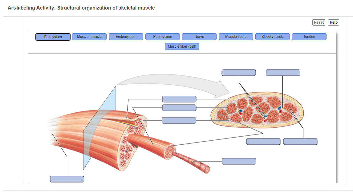
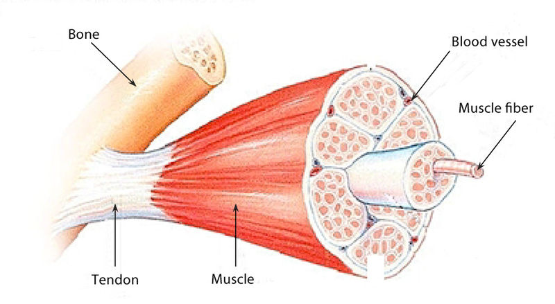
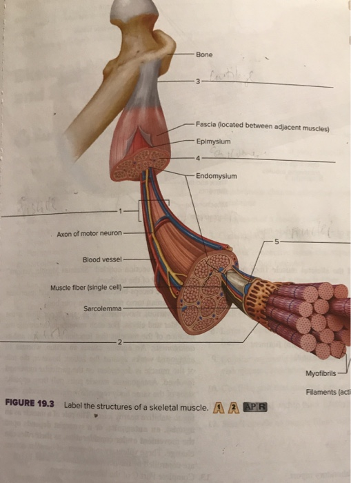
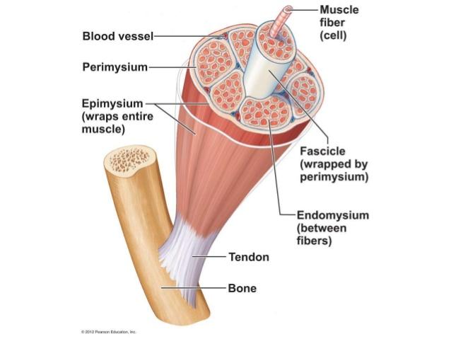
Label the structures of a skeletal muscle fiber. Structure of Skeletal Muscle | SEER Training Each skeletal muscle fiber is a single cylindrical muscle cell. An individual skeletal muscle may be made up of hundreds, or even thousands, of muscle fibers bundled together and wrapped in a connective tissue covering. Each muscle is surrounded by a connective tissue sheath called the epimysium. 10.2 Skeletal Muscle - Anatomy and Physiology 2e | OpenStax Each skeletal muscle is an organ that consists of various integrated tissues. These tissues include the skeletal muscle fibers, blood vessels, nerve fibers, and connective tissue. Each skeletal muscle has three layers of connective tissue (called "mysia") that enclose it and provide structure to the muscle as a whole, and also ... muscle fiber diagram labeled BIO201-Muscle Fiber | Human Anatomy And Physiology, Muscle Anatomy . muscle fiber model anatomy cell human neuron synaptic models motor labeled physiology body skeletal ranvier muscles mitochondria node cleft junction. Figure 1114 - Body Function - 78 Steps Health . figure muscle fiber function anatomy 1114 ... Muscle and Nervous Tissue Review Flashcards | Quizlet Place the organizational level of muscle tissue in order, beginning with the entire muscle and ending with the smallest component. 1.) Muscle 2.) Fascicle 3.) Muscle Fiber 4.) Myofibril 5.) Myofilament Label the components of skeletal muscle. Label the connective tissue in the figure.
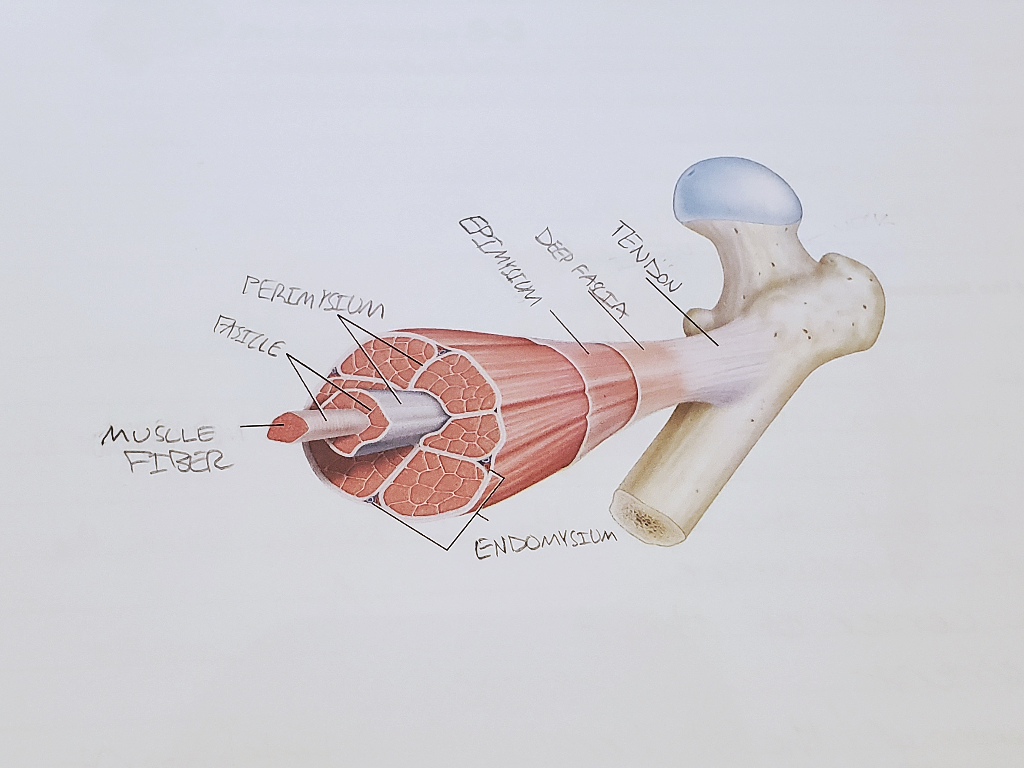
Skeletal Muscle | Anatomy and Physiology I - Lumen Learning Bundles of muscle fibers, called fascicles, are covered by the perimysium. Muscle fibers are covered by the endomysium. Each skeletal muscle is an organ that consists of various integrated tissues. These tissues include the skeletal muscle fibers, blood vessels, nerve fibers, and connective tissue. 10.2 Skeletal Muscle - Anatomy & Physiology Figure 10.2.1 - The Three Connective Tissue Layers: Bundles of muscle fibers, called fascicles, are covered by the perimysium. Muscle fibers are covered by the endomysium. Inside each skeletal muscle, muscle fibers are organized into bundles, called fascicles, surrounded by a middle layer of connective tissue called the perimysium. 9.2A: Skeletal Muscle Fibers - Medicine LibreTexts Skeletal muscles are composed of striated subunits called sarcomeres, which are composed of the myofilaments actin and myosin. Learning Objectives Outline the structure of a skeletal muscle fiber Key Points Muscles are composed of long bundles of myocytes or muscle fibers. Myocytes contain thousands of myofibrils. structure of skeletal muscle fiber Flashcards | Quizlet covers the entire skeletal muscle Perimysium The connective tissue that surrounds fascicles. Endomysium Surrounds individual muscle fibers Sarcolemma plasma membrane of a muscle fiber Sarcoplasm cytoplasm of a muscle cell sarcoplasmic reticulum specialized endoplasmic reticulum of muscle cells terminal cisternae
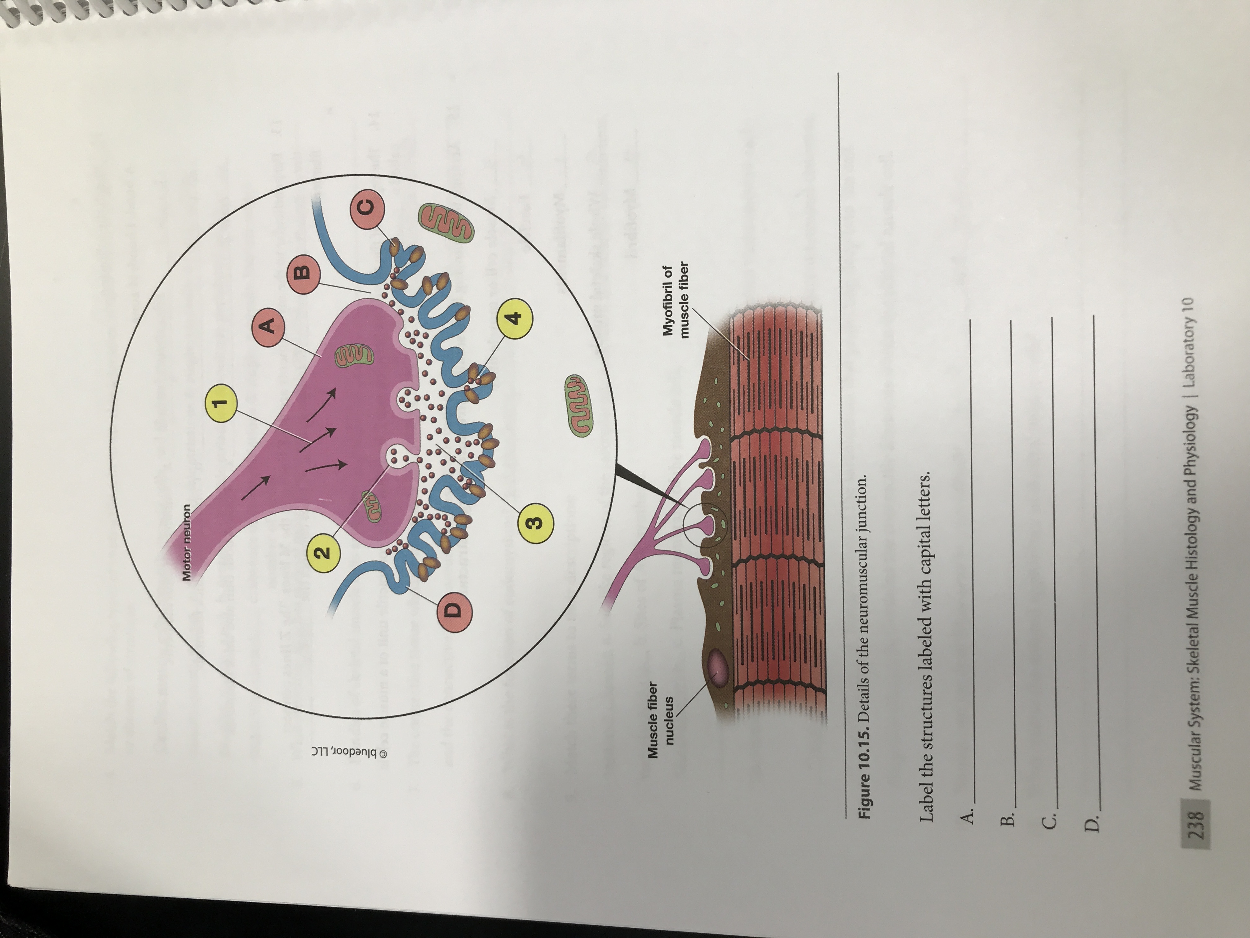
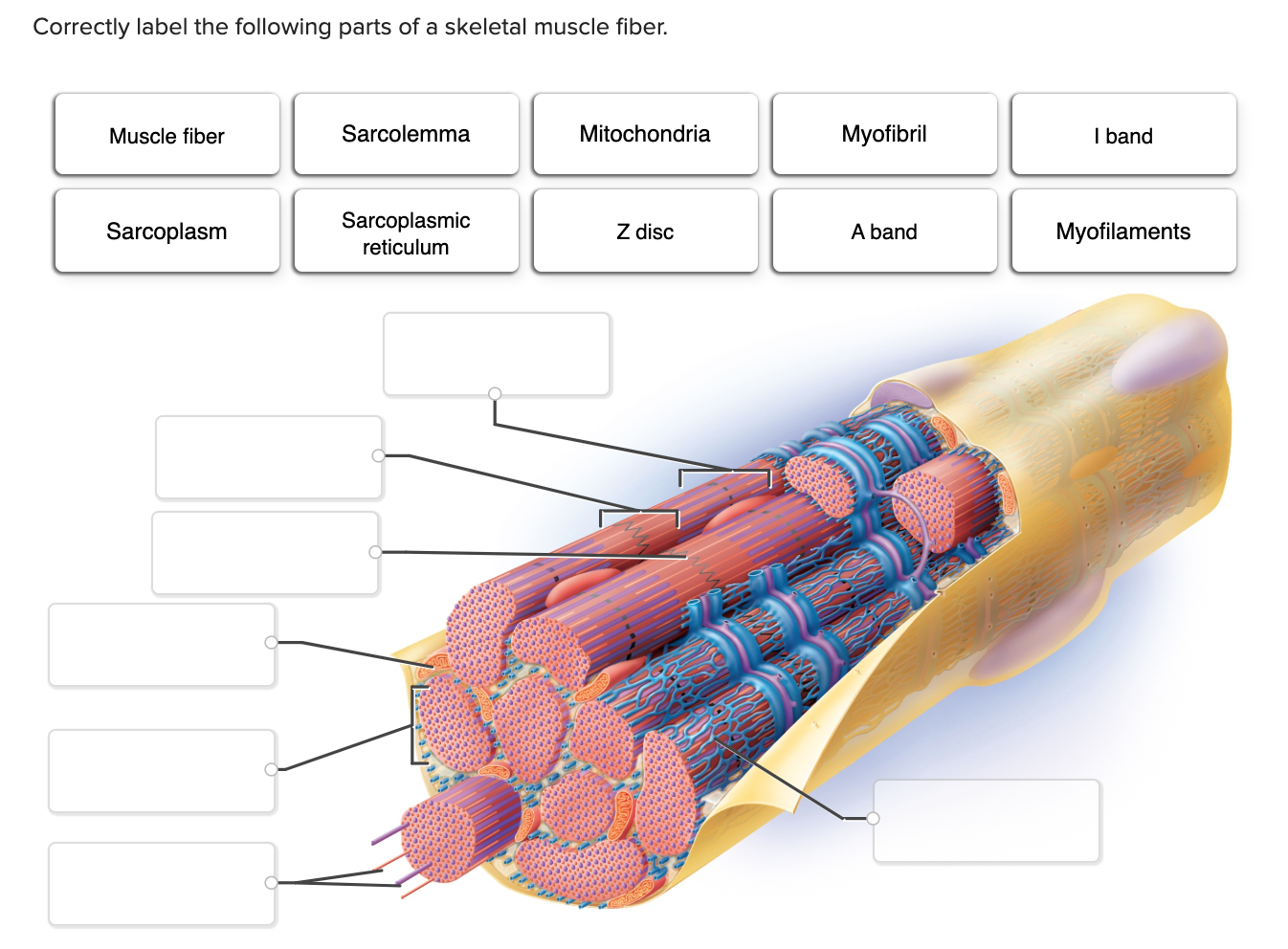
Skeletal muscle fibers: arrangement and diagram | GetBodySmart Skeletal muscle fibers are located inside muscles, where they are organized into bundles called fascicles (= fasciculi). 1 2 3 The epimysium is the connective tissue layer that covers the outer surface of the muscle. 1 2 Surrounding and holding together each fascicle is a layer of connective tissue known as perimysium. Neuromuscular junction: Parts, structure and steps | Kenhub At its simplest, the neuromuscular junction is a type of synapse where neuronal signals from the brain or spinal cord interact with skeletal muscle fibers, causing them to contract. The activation of many muscle fibers together causes muscles to contract, which in turn can produce movement. The neuromuscular junction then, is a key component in ... skeletal muscle label anatomy 30 Label The Structures Of A Skeletal Muscle - Labels For Your Ideas opilizeb.blogspot.com. skeletal structure fibers function fascicle quizlet myofibrils endomysium perimysium exercising fascicles contraction fascia myofibril surrounded exercise labeling 1001. Correctly label the following parts of a skeletal muscle fiber ... Science Anatomy and Physiology Correctly label the following parts of a skeletal muscle fiber. Mitochondria Sarcolemma Sarcoplasmic reticulum Openings into transverse tubules Triad: Terminal cisterns Muscle fiber Sarcoplasm Triad: Transverse tubule Myofibril Nucleus. Correctly label the following parts of a skeletal muscle fiber. Mitochondria ...
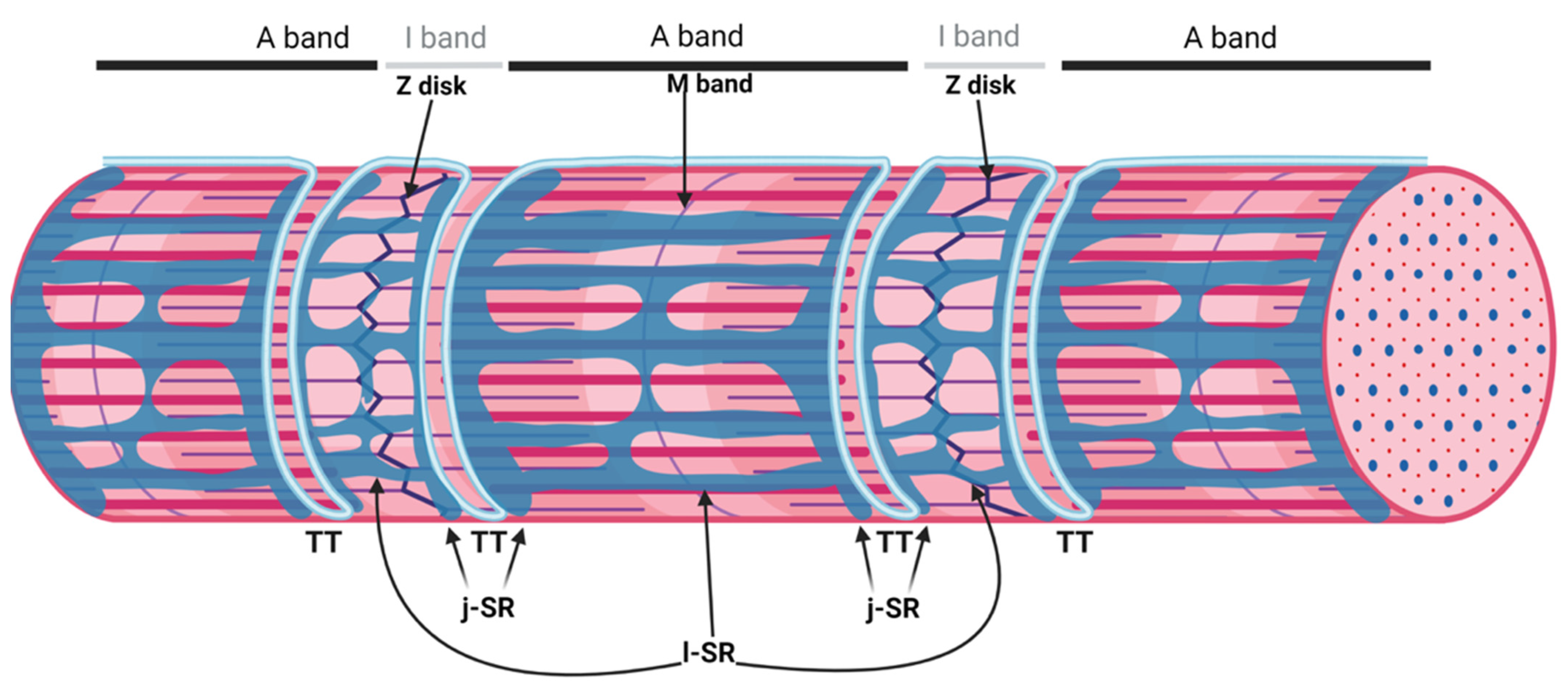
Skeletal muscle tissue: Histology | Kenhub Special terms are used to describe structures associated with skeletal muscle tissue. Muscle tissue terms often begin with myo-, mys-, or sarco-. The cytoplasm of a muscle cells is referred to as sarcoplasm.The plasma membrane is called the sarcolemma and the endoplasmic reticulum is called the sarcoplasmic reticulum.A muscle fiber may also be referred to as a myofiber.
Skeletal Muscle Fiber Structure and Function - Open Textbooks for Hong Kong The striated appearance of skeletal muscle tissue is a result of repeating bands of the proteins actin and myosin that occur along the length of myofibrils. Myofibrils are composed of smaller structures called myofilaments. There are two main types of myofilaments: thick filaments and thin filaments.
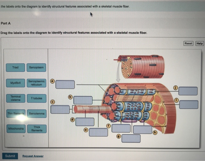
Structures of the Skeletal Muscle Fiber Flashcards | Quizlet -Muscle fibers are filled with threads called myofibrils separated by SR (sarcoplasmic reticulum) -Myofilaments (thick & thin filaments) are the contractile proteins of muscle that actually cause muscles to contract. Sarcoplasmic Reticulum (SR) -Stores Ca+2 in a relaxed muscle -Release of Ca+2 triggers muscle contraction Filaments and the Sarcomere
Skeletal Muscle Fiber - GetBodySmart Skeletal Muscle Fiber. Skeletal muscles are the types of muscle tissue that enable us with voluntary movements. They are attached to the bones of the skeleton by tendons. Skeletal muscle fibers have a striated (striped) appearance on histological sections because they are made up of smaller units called sarcomeres that run parallel to each other, giving the muscle the striated appearance.
Solved Internal Structure of a Skeletal Muscle Cell Figure | Chegg.com Question: Internal Structure of a Skeletal Muscle Cell Figure 10.8. Label this diagram using the descriptions below. Muscle fibers: Alternative name for skeletal muscle cells. Nucleus: Contains the genetic material. • Sarcolemma: Plasma membrane of the muscle cell. • Sarcoplasmic reticulum (SR): Interconnecting tubules of endoplasmic ...
11.2: Microscopic Anatomy of Skeletal Muscles - Biology LibreTexts Skeletal muscle is found attached to bones. It consists of long multinucleate fibers. The fibers run the entire length of the muscle they come from and so are usually too long to have their ends visible when viewed under the microscope. The fibers are relatively wide and very long, but unbranched.
Skeletal Muscle Histology Slide Identification and Labeled Diagram ... From the skeletal muscle histology slide, you might identify the following important structures under the light microscope. Please try to find out these structures from the skeletal muscle slide labeled images. #1. Longitudinal section of skeletal muscle #2. Cross-section of skeletal muscle #3. Skeletal muscle fibers of the longitudinal section #3.
10.3 Muscle Fiber Contraction and Relaxation - OpenStax Relaxation of a Skeletal Muscle. Relaxing skeletal muscle fibers, and ultimately, the skeletal muscle, begins with the motor neuron, which stops releasing its chemical signal, ACh, into the synapse at the NMJ. The muscle fiber will repolarize, which closes the gates in the SR where Ca ++ was being released. ATP-driven pumps will move Ca ++ out ...
Solved Muscle Cell Label the structures of a skeletal muscle - Chegg Muscle Cell Label the structures of a skeletal muscle fiber. Nucleus Myofibril Sarcolemma Sarcoplasmic reticulum Openings into T tubules < Prev 3 of 15 !!! Next > Thinkinys - How to write a boty The Good Cre. Dob C ommunicatio pdf Communication.pdf This problem has been solved!
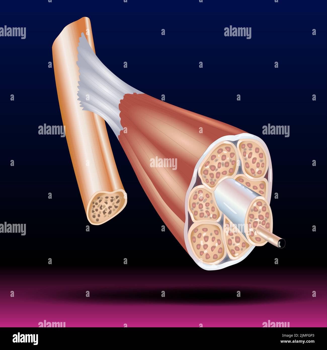
Skeletal Muscle: Definition, Function, Structure, Location | Biology ... Skeletal muscle is comprised of a series of muscle fibers made of muscle cells. These muscle cells are long and multinucleated. At the ends of each skeletal muscle a tendon connects the muscle to bone. This tendon connects directly to the epimysium, or collagenous outer covering of skeletal muscle. Underneath the epimysium, muscle fibers are ...
skeletal muscle label anatomy 37 Label The Structures Of A Skeletal Muscle - Labels 2021. 12 Images about 37 Label The Structures Of A Skeletal Muscle - Labels 2021 : 13 Best Images of Muscle Labeling Worksheet - Label Muscles Worksheet, Simple Muscle Anatomy Simple Muscle Diagram For Kids Label The Major and also 37 Label The Structures Of A Skeletal Muscle - Labels 2021.
IJMS | Free Full-Text | Comprehensive Analysis of Long Noncoding RNA ... Skeletal muscle is a heterogeneous tissue, composed of different types of muscle fibers, with distinct contractile and metabolic characteristics [].Myosin heavy chain (MyHC) is the major contractile protein of skeletal muscle cells [].According to the electrophoretic analysis results of myosin heavy chain isoforms in adult mammals, muscle fibers are mainly divided into type I (MyHC I) and type ...


















Post a Comment for "45 label the structures of a skeletal muscle fiber"