43 diagram of a light microscope with labels
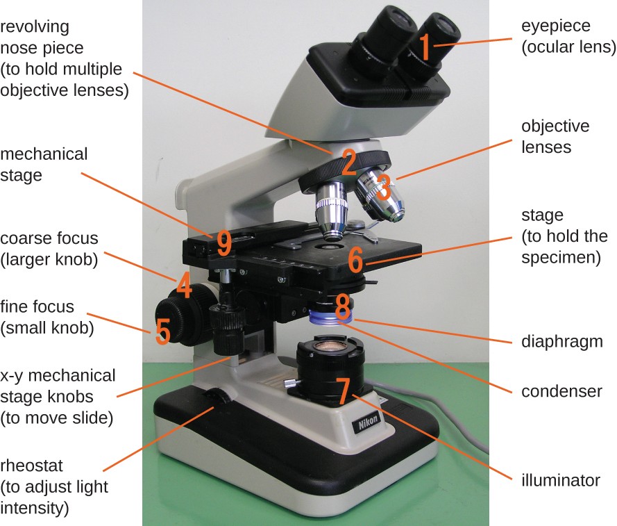
Light Microscope-Definition, Principle, Types, Parts, Labeled Diagram ... A light microscope is a device or instrument used in biology laboratories that uses visible light to locate, magnify, and expand micro objects. Using lenses, they focus light on the specimen and magnify it to generate a photograph. Typically, the specimen is positioned close to the microscopic lens. Microscope Diagram Labeled, Unlabeled and Blank | Parts of a Microscope 10. Condenser - Focuses light from the light source onto the specimen. 11. Iris Diaphragm - An opaque iris composed of blades made to pass light through an aperture. 12. Base - The supporting block of the light microscope. 13. Light Source - A light or a daylight directed via a mirror. 14.
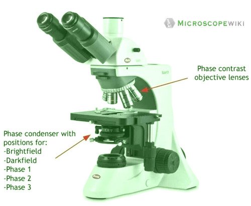
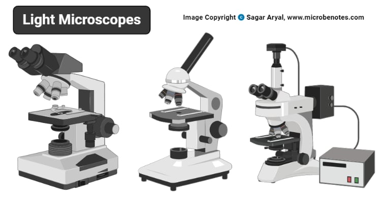
Light Microscope Labeled Diagram, Definition, Principle, Types, Parts ... Light Microscope Labeled Diagram - Principle of Brightfield Microscope Parts of a bright-field microscope or Compound light microscope An optical microscope, the bright-field microscope (or compound light microscope) is an invaluable tool in the fields of biology, medicine, and education.
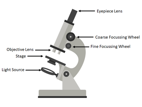
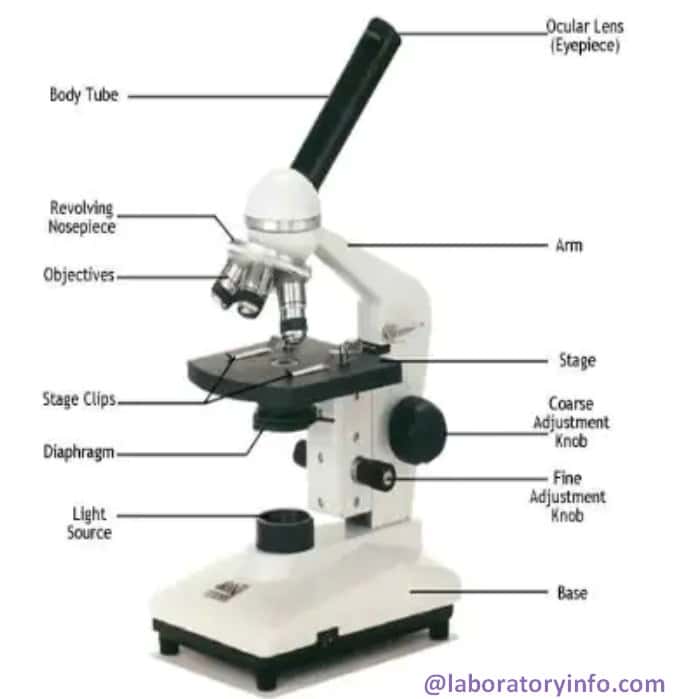
Diagram of a light microscope with labels
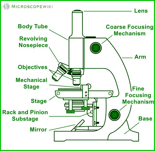
Parts of a microscope with functions and labeled diagram - Microbe Notes Figure: Diagram of parts of a microscope There are three structural parts of the microscope i.e. head, base, and arm. Head - This is also known as the body. It carries the optical parts in the upper part of the microscope. Base - It acts as microscopes support. It also carries microscopic illuminators. Label the microscope — Science Learning Hub Use this interactive to identify and label the main parts of a microscope. Drag and drop the text labels onto the microscope diagram. diaphragm or iris base eye piece lens fine focus adjustment light source stage coarse focus adjustment high-power objective Download Exercise Microscopy: Intro to microscopes & how they work (article) - Khan Academy In most cases, the part of a cell or tissue that we want to look at isn't naturally fluorescent, and instead must be labeled with a fluorescent dye or tag before it goes on the microscope. The leaf picture at the start of the article was taken using a specialized kind of fluorescence microscopy called confocal microscopy.
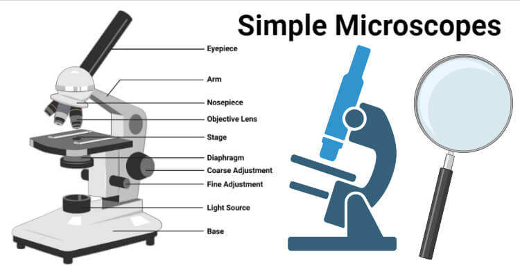
Diagram of a light microscope with labels. Compound Microscope Parts, Functions, and Labeled Diagram Compound Microscope Definitions for Labels Eyepiece (ocular lens) with or without Pointer: The part that is looked through at the top of the compound microscope. Eyepieces typically have a magnification between 5x & 30x. Monocular or Binocular Head: Structural support that holds & connects the eyepieces to the objective lenses. Microscope Types (with labeled diagrams) and Functions Electron microscope labeled diagram The different types of electron microscopes are: Transmission Electron Microscope Scanning Electron Microscope Reflection Electron Microscope Scanning transmission electron microscope Scanning tunneling microscopy Electron microscope functions: Semiconductors and Data Storage Industry Failure Analysis Parts of the Microscope (Labeled Diagrams) Simple microscope labelled diagram Image created with Biorender Tube/Body Tube It serves as the connector between the eyepiece/ocular and objective lenses. Objective lenses The lenses have varying magnifying power, which typically consists of 10x, 40x, and 100x. Microscope Parts and Functions This allows the slide to be easily inserted or removed from the microscope. It also allows the specimen to be labeled, transported, and stored without damage. Stage: The flat platform where the slide is placed. Stage clips: Metal clips that hold the slide in place.
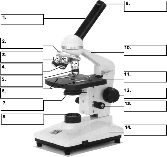
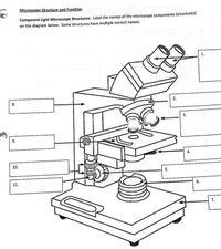
Light microscopes - Cell structure - Edexcel - BBC Bitesize The magnification of a lens is shown by a multiplication sign followed by the amount the lens magnifies. So a lens magnifying ten times would be ×10. The total magnification of a microscope is:... Microscope Parts, Types & Diagram | What is a Microscope? There are many illustrations available for the diagram of a light microscope. The essential parts include the head, base, arms, lenses, and lights. In diagrams, one would see the head... Microscope Labeling - The Biology Corner Microscope Labeling. Shannan Muskopf May 31, 2018. This simple worksheet pairs with a lesson on the light microscope, where beginning biology students learn the parts of the light microscope and the steps needed to focus a slide under high power. The labeling worksheet could be used as a quiz or as part of direct instruction where students ... 1.5: Microscopy - Biology LibreTexts In Biology, the compound light microscope is a useful tool for studying small specimens that are not visible to the naked eye. The microscope uses bright light to illuminate through the specimen and provides an inverted image at high magnification and resolution. ... Blank microscope to label parts. This page titled 1.5: Microscopy is shared ...
A Study of the Microscope and its Functions With a Labeled Diagram ... To better understand the structure and function of a microscope, we need to take a look at the labeled microscope diagrams of the compound and electron microscope. These diagrams clearly explain the functioning of the microscopes along with their respective parts. Man's curiosity has led to great inventions. The microscope is one of them. PDF Parts of the Light Microscope - Science Spot Supports the MICROSCOPE D. STAGE CLIPS HOLD the slide in place C. OBJECTIVE LENSES Magnification ranges from 10 X to 40 X F. LIGHT SOURCE Projects light UPWARDS through the diaphragm, the SPECIMEN, and the LENSES H. DIAPHRAGM Regulates the amount of LIGHT on the specimen E. STAGE Supports the SLIDE being viewed K. ARM Used to SUPPORT the Light Microscope- Definition, Principle, Types, Parts, Labeled Diagram ... Figure: Labeled Diagram of a Light Microscope. Types of light microscopes (optical microscope) With the evolved field of Microbiology, the microscopes used to view specimens are both simple and compound light microscopes, all using lenses. Binocular Microscope Anatomy - Parts and Functions with a Labeled Diagram The important non-optical parts of the light compound microscope are the body tube or head, arm or frame, fine adjustment, coarse adjustment, nose piece, stage, and base. Now, I will describe all these non-optical parts of the light compound microscope with the labeled diagrams. The body tube of the microscope
Compound Microscope - Diagram (Parts labelled), Principle and Uses A compound microscope: Is used to view samples that are not visible to the naked eye. Uses two types of lenses - Objective and ocular lenses. Has a higher level of magnification - Typically up to 2000x. Is used in hospitals and forensic labs by scientists, biologists and researchers to study microorganisms. Invented in the late 16th century ...
Compound Microscope Parts - Labeled Diagram and their Functions Labeled diagram of a compound microscope Major structural parts of a compound microscope There are three major structural parts of a compound microscope. The head includes the upper part of the microscope, which houses the most critical optical components, and the eyepiece tube of the microscope.
Microscopy: Intro to microscopes & how they work (article) - Khan Academy In most cases, the part of a cell or tissue that we want to look at isn't naturally fluorescent, and instead must be labeled with a fluorescent dye or tag before it goes on the microscope. The leaf picture at the start of the article was taken using a specialized kind of fluorescence microscopy called confocal microscopy.
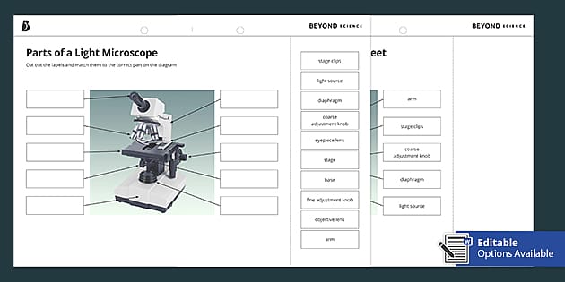
Label the microscope — Science Learning Hub Use this interactive to identify and label the main parts of a microscope. Drag and drop the text labels onto the microscope diagram. diaphragm or iris base eye piece lens fine focus adjustment light source stage coarse focus adjustment high-power objective Download Exercise
Parts of a microscope with functions and labeled diagram - Microbe Notes Figure: Diagram of parts of a microscope There are three structural parts of the microscope i.e. head, base, and arm. Head - This is also known as the body. It carries the optical parts in the upper part of the microscope. Base - It acts as microscopes support. It also carries microscopic illuminators.

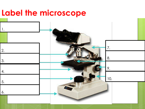

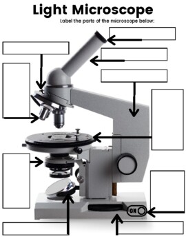











![What is Compound Microscope? - Diagram, Function [updated]](https://www.tutoroot.com/blog/wp-content/uploads/2022/09/Compound-microscope-1.png)


Post a Comment for "43 diagram of a light microscope with labels"