42 label the transmission electron micrograph of the mitochondrion.
Solved Label the transmission electron micrograph of the 100% (4 ratings) Explanation - Mitochondrion is filamentous or globular in shape, occur in variable numbers from a few hundred to few thousands in different cells. It …. View the full answer. Transcribed image text: Label the transmission electron micrograph of the mitochondrion. Matrix granule Mitochondrion Outer membrane Cristae Inner ... Frontiers | Mitochondrial Morphology and Mitophagy in Heart Diseases ... Transmission electron microscopy (TEM) has long been an important technique, capable of high degree resolution and visualization of subcellular structures and organization. Over the last 20 years, TEM has gained popularity in the cardiovascular field to visualize changes at the nanometer scale in cardiac ultrastructure during cardiovascular development, aging, and a broad range of pathologies.
Brock Biology of Microorganisms 13th Edition - academia.edu Enter the email address you signed up with and we'll email you a reset link.

Label the transmission electron micrograph of the mitochondrion.
Mitochondria under the microscope - Science Learning Hub Microscopes have been crucial for our understanding of mitochondrial structure and function. Mitochondria are visible under the light microscope although little detail can be seen. Transmission electron microscopy (left) shows the complex internal membrane structure of mitochondria, and electron tomography (right) gives a three-dimensional view. PDF Fast Transmission Electron Microscope (TEM) Technique for ... - Longdom Herbicides; Fast Transmission Electron Microscope (TEM); Human and animal mitochondria. Introduction. The present article is based on the thesis: Some effects of low . doses of simazine and bentazone on fine structure and metabolism of soybean cells [1-3]. The great similarity existing among plant, human Transmission Electron Microscopy for Analysis of Mitochondria in Mouse ... Transmission electron microscopy (TEM) is a powerful technique for ultrastructural studies ( Watson, 1958 ). TEM has been very useful in studying mitochondrial structure in skeletal muscle in both physiological and pathological conditions ( Picard et al., 2013 ).
Label the transmission electron micrograph of the mitochondrion.. PDF Multi-color live-cell STED nanoscopy of mitochondria with a gentle ... 24 Abstract 25 Capturing mitochondria's intricate and dynamic structure poses a daunting challenge for optical nanoscopy. 26 Different labeling strategies have been demonstrated for live-cell stimulated emission depletion (STED) 27 microscopy of mitochondria, but orthogonal strategies are yet to be established, and image acquisition has 28 suffered either from photodamage to the organelles or ... PDF Identifying Organelles from an Electron Micrograph ELECTRON MICROGRAPH OF MITOCHONDRION Courtesy of Electron Microscopy Unit University of Lancaster endoplasmic cristae reticulum fluid matrix outer membrane ER, cristae, fluid matrix, ribosome, outer membrane Courtesy of Dr. Julian Thorpe - EM & FACS Lab, Biological Sciences University Of Sussex Bio 232 ~ Lab Midterm Flashcards | Quizlet Label the transmission electron micrograph of the mitochondrion. Label the membranous organelles. Place the following cytoplasmic structures in the appropriate structural category. Analysis of Yeast Mitochondria by Electron Microscopy We provide here detailed protocols for the analysis of yeast mitochondria by transmission electron microscopy: (1) chemical fixation and Epon embedding of yeast cells and isolated mitochondria, and (2) cryosectioning and immunolabeling of yeast cells and isolated mitochondria according to the Tokuyasu method. less
IB Questionbank Test | Ankit Mistry - Academia.edu It lacks a nucleus. B. It lacks a cell wall. C. It has only one mitochondrion. D. It lacks subdivision into separate cells. 37. [1 mark] The image shows an electron micrograph of mesophyll cells. What is the name of the structure labelled X? A. Cytoplasm B. Mitochondrion C. Nucleus D. Chloroplast 39. Mitochondria and Endoplasmic Reticulum Imaging by Correlative Light and ... Transmission electron microscopy provides good membrane contrast and nanometer-scale resolution for the observation of cellular organelles; however, it is exceptionally time-consuming when assessing the three-dimensional (3D) structure of highly curved organelles. Labeling the Cell Flashcards | Quizlet Label the transmission electron micrograph of the mitochondrion. Label the transmission electron micrograph of the nucleus. membrane bound organelles golgi apparatus, mitochondrion, lysosome, peroxisome, rough endoplasmic reticulum nonmembrane bound organelles ribosomes, centrosome, proteasomes cytoskeleton includes Transmission electron microscopy techniques - University of Otago STEM (scanning transmission electron microscopy) enables the use of other of signals that cannot be spatially correlated in TEM, including secondary electrons, scattered beam electrons, characteristic X-rays, and electron energy loss—allowing very accurate elemental mapping and chemical analysis of samples. Single Particle Reconstruction
Transmission Electron Microscopy - an overview | ScienceDirect Topics platelet transmission electron microscopy (tem) is an essential tool for laboratory diagnosis of various hereditary platelet disorders since it was first used to visualize fibrin-platelet clot formation in 1950s.51,52 platelet tem employs two main methods to visualize platelet ultrastructure, whole-mount tem and thin-section tem. 53,54 … Electron Micrographs of Cell Organelles - Biology Discussion It is an electron micrograph of cell's largest and most important organelle - the mitochondria and is characterized by the following features (Fig. 7 & 8): (1) The name mitochondria was given by Benda (1898) and their ma n function was brought to light by Kingsbury (1912). cells part 1.pdf - 3 0 1 . 3 Figure 2 shows a photograph ... - Course Hero * 03 * Turn over IB/M/Jun18/7401/1 Do not write outside the box Figure 2 shows a photograph of part of a mitochondrion from a mouse liver cell taken using a transmission electron microscope at × 62 800 magnification. Figure 2 Produce a scientific drawing of the mitochondrion in Figure 2 in the box below. Recent structural insight into mitochondria gained by microscopy Novel applications of microscopy have recently provided new insights into mitochondrial structures. Diverse techniques such as high resolution scanning electron microscopy, transmission electron microscopy, electron microscope tomography and light microscopy have contributed a better understanding of mitochondrial compartmentalization, dynamic networks of mitochondria, intermembrane bridges ...
Mitochondrion,TEM[01808024644]| 写真素材・ストックフォト・画像・イラスト素材|アマナイメージズ
Cambridge International AS and A Level Biology Coursebook … Cambridge International AS and A Level Biology Coursebook Fourth Edition

Biology Club: Our cells #1 - ( structure, function, division, disorder ...
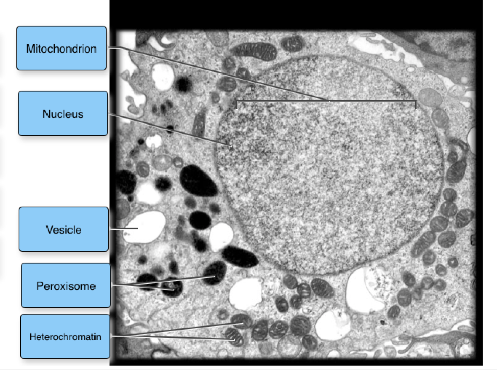
Solved Label the transmission electron micrograph of the - Chegg Question: Label the transmission electron micrograph of the cell. 0 Nucleus rences Mitochondrion Heterochromatin Peroxisome Vesicle ULAR bumit Click and drag each label into the correct category to indicate whether it pertains to the cytoplasm or the plasma membrane.

Cell Types and Organelles
Microbiology Module 3 Flashcards - Quizlet The internal compartment is comprised of the cytoplasm, the nucleus, cytoskeletal components, and other organelles, such as mitochondria and ribosomes. Label the image to test your knowledge of eukaryotic cell structure and function. Flagellum organelle used for locomotion Golgi apparatus site of protein modification + lysosome formation

What do cristae do for mitochondria? - proquestyamaha.web.fc2.com
Transmission electron microscopy of C. parvum sporozoites showing the ... The ribosome- studded mitochondrion ( ء ) is between the nucleus (N) and the crys- talloid body (CB). The apical organelles shown include the mi- cronemes (M) and dense granules (D) for entry into...
Solved: Please Answer Part A And B For The Animal Cell And... | Chegg.com
Mitochondrial morphology and function: two for the price of one! This work represents a technical advance that allows the correlation of mitochondrial function and morphology with greater resolution and volume than has previously been feasible. LAY SUMMARY: Transmission electron microscopy (TEM) is a high-resolution technique used for the study of cells and their components, such as mitochondria.
33 Label The Transmission Electron Micrograph Of The Nucleus - Label ...
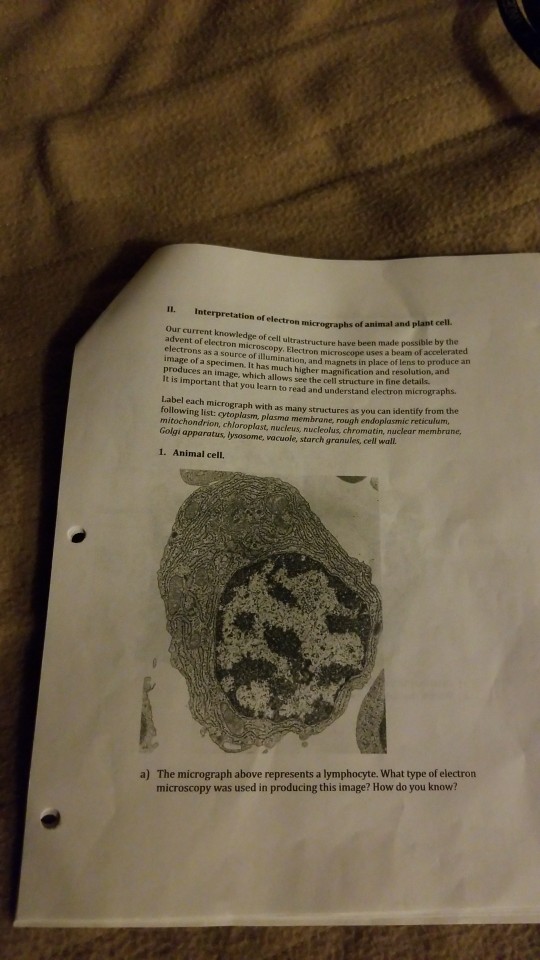
Apoptosis: A Review of Programmed Cell Death - PMC Figure 2A is a transmission electron micrograph (TEM) of the normal thymus tissue depicted in Figure 1C. The lymphocytes are closely packed, have large nuclei and scant cytoplasm. Figure 2B is a TEM of apoptotic thymic lymphocytes in an early phase of apoptosis with condensed and peripheralized chromatin. The cytoplasm is beginning to condense ...
Post a Comment for "42 label the transmission electron micrograph of the mitochondrion."